Explore
 Validate
 Learn
Learn Western blot
Antibody data
- Antibody Data
- Antigen structure
- References [0]
- Comments [0]
- Validations
- Western blot [4]
- Immunohistochemistry [1]
Submit
Validation data
Reference
Comment
Report error
- Product number
- PA5-22149 - Provider product page

- Provider
- Invitrogen Antibodies
- Product name
- MX1 Polyclonal Antibody
- Antibody type
- Polyclonal
- Antigen
- Recombinant protein fragment
- Description
- Recommended positive controls: NCI-H929, human MX1-transfected 293T.
- Concentration
- 0.06 mg/mL
No comments: Submit comment
Supportive validation
- Submitted by
- Invitrogen Antibodies (provider)
- Main image
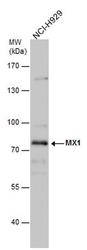
- Experimental details
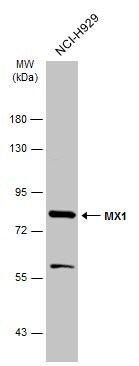
- Western blot analysis of MX1 in whole cell extracts (30 µg). Samples was separated by 7.5% SDS-PAGE and the membrane was probed with MX1 Polyclonal antibody (Product # PA5-22149) at a dilution of 1:1000.
- Submitted by
- Invitrogen Antibodies (provider)
- Main image
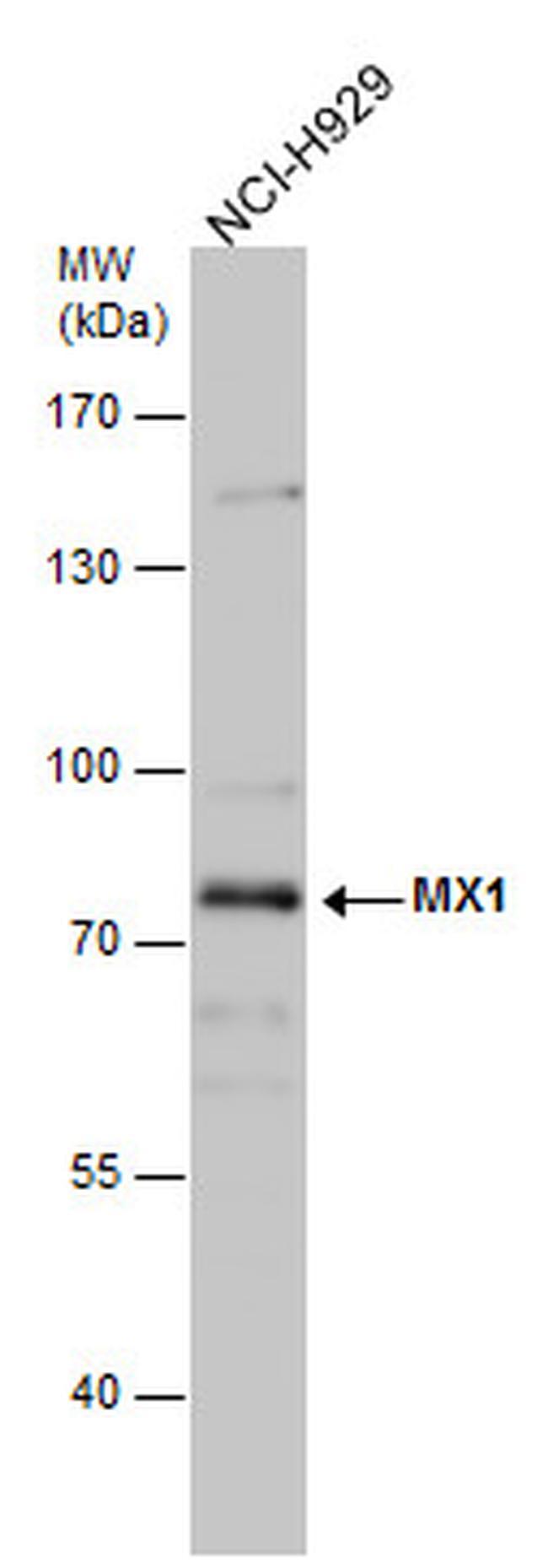
- Experimental details
- Western Blot analysis of MX1 was performed by separating 30 µg of Whole cell extracts by 7.5% SDS-PAGE. Proteins were transferred to a membrane and probed with a MX1 Polyclonal Antibody (Product # PA5-22149) at a dilution of 1:1000. The HRP-conjugated anti-rabbit IgG antibody was used to detect the primary antibody.
- Submitted by
- Invitrogen Antibodies (provider)
- Main image
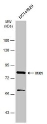
- Experimental details
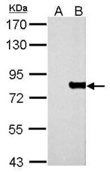
- MX1 Polyclonal Antibody detects MX1 protein by western blot analysis. A. 30 µg 293T whole cell lysate/extract. B. 30 µg whole cell lysate/extract of human MX1-transfected 293T cells.7.5% SDS-PAGE. MX1 Polyclonal Antibody (Product # PA5-22149) dilution: 1:5,000. The HRP-conjugated anti-rabbit IgG antibody was used to detect the primary antibody.

- Submitted by
- Invitrogen Antibodies (provider)
- Main image
- Experimental details
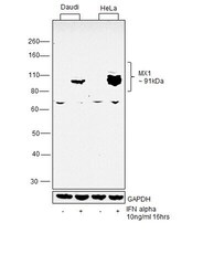
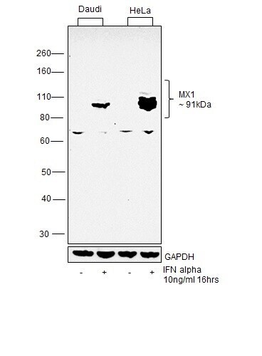
- Western blot was performed using MX1 Polyclonal Antibody (Product # PA5-22149) and a 91 kDa band corresponding to MX1 was observed in cell lines treated with IFN alpha. Whole cell extracts (30 µg lysate) of Daudi (Lane 1), Daudi treated with IFN Alpha (10ng/mL, 16 hrs) (Lane 2), HeLa (Lane 3) and HeLa treated with IFN Alpha (10ng/mL, 16 hrs) were electrophoresed using NuPAGE® 4-12 % Bis-Tris gel (Product # NP0322BOX). Resolved proteins were then transferred onto a nitrocellulose membrane (Product # IB23001) by iBlot® 2 Dry Blotting System (Product # IB21001). The blot was probed with the primary antibody (1:5000 dilution) and detected by chemiluminescence with Goat anti-Rabbit IgG (H+L), Superclonal™ Recombinant Secondary Antibody, HRP (Product # A27036, 1:4000 dilution) using the iBright FL 1000 (Product # A32752). Chemiluminescent detection was performed using Novex® ECL Chemiluminescent Substrate Reagent Kit (Product # WP20005).
Supportive validation
- Submitted by
- Invitrogen Antibodies (provider)
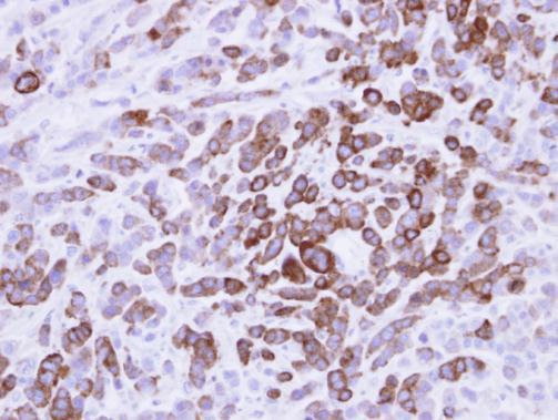
- Main image

- Experimental details
- MX1 Antobody detects MX1 protein at cytosol on MDA-MB-468 xenograft by immunohistochemical analysis. Sample: Paraffin-embedded MDA-MB-468 xenograft. MX1 Antobody (Product # PA5-22149) dilution: 1:500. Antigen Retrieval: EDTA based buffer, pH 8.0, 15 min.