Explore
 Validate
 Learn
LearnPA5-23316
antibody from Invitrogen Antibodies
Targeting: IL1RL1
DER4, FIT-1, IL33R, ST2, ST2L, ST2V, T1
 Western blot
Antibody data
- Antibody Data
- Antigen structure
- References [5]
- Comments [0]
- Validations
- Western blot [3]
- Immunohistochemistry [1]
- Other assay [2]
Submit
Validation data
Reference
Comment
Report error
- Product number
- PA5-23316 - Provider product page

- Provider
- Invitrogen Antibodies
- Product name
- ST2 Polyclonal Antibody
- Antibody type
- Polyclonal
- Antigen
- Other
- Reactivity
- Human, Mouse, Rat
- Host
- Rabbit
- Isotype
- IgG
- Vial size
- 100 µg
- Concentration
- 1.0 mg/mL
- Storage
- Store at 4°C short term. For long term storage, store at -20°C, avoiding freeze/thaw cycles.
Submitted references Hyperactivity of Innate Immunity Triggers Pain via TLR2-IL-33-Mediated Neuroimmune Crosstalk.
Dynamic Expression of Interleukin-33 and ST2 in the Mouse Reproductive Tract Is Influenced by Superovulation.
Identification of TNFR2 and IL-33 as therapeutic targets in localized fibrosis.
IL-33/ST2 signaling excites sensory neurons and mediates itch response in a mouse model of poison ivy contact allergy.
Spinal IL-33/ST2 Signaling Contributes to Neuropathic Pain via Neuronal CaMKII-CREB and Astroglial JAK2-STAT3 Cascades in Mice.
Huang J, Gandini MA, Chen L, M'Dahoma S, Stemkowski PL, Chung H, Muruve DA, Zamponi GW
Cell reports 2020 Oct 6;33(1):108233
Cell reports 2020 Oct 6;33(1):108233
Dynamic Expression of Interleukin-33 and ST2 in the Mouse Reproductive Tract Is Influenced by Superovulation.
Begum S, Perlman BE, Valero-Pacheco N, O'Besso V, Wu T, Morelli SS, Beaulieu AM, Douglas NC
The journal of histochemistry and cytochemistry : official journal of the Histochemistry Society 2020 Apr;68(4):253-267
The journal of histochemistry and cytochemistry : official journal of the Histochemistry Society 2020 Apr;68(4):253-267
Identification of TNFR2 and IL-33 as therapeutic targets in localized fibrosis.
Izadi D, Layton TB, Williams L, McCann F, Cabrita M, Espirito Santo AI, Xie W, Fritzsche M, Colin-York H, Feldmann M, Midwood KS, Nanchahal J
Science advances 2019 Dec;5(12):eaay0370
Science advances 2019 Dec;5(12):eaay0370
IL-33/ST2 signaling excites sensory neurons and mediates itch response in a mouse model of poison ivy contact allergy.
Liu B, Tai Y, Achanta S, Kaelberer MM, Caceres AI, Shao X, Fang J, Jordt SE
Proceedings of the National Academy of Sciences of the United States of America 2016 Nov 22;113(47):E7572-E7579
Proceedings of the National Academy of Sciences of the United States of America 2016 Nov 22;113(47):E7572-E7579
Spinal IL-33/ST2 Signaling Contributes to Neuropathic Pain via Neuronal CaMKII-CREB and Astroglial JAK2-STAT3 Cascades in Mice.
Liu S, Mi WL, Li Q, Zhang MT, Han P, Hu S, Mao-Ying QL, Wang YQ
Anesthesiology 2015 Nov;123(5):1154-69
Anesthesiology 2015 Nov;123(5):1154-69
No comments: Submit comment
Supportive validation
- Submitted by
- Invitrogen Antibodies (provider)
- Main image

- Experimental details
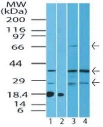
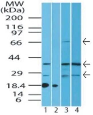
- Western blot analysis of ST2 protein in human kidney lysate in the 1) absence and 2) presence of immunizing peptide, 3) mouse kidney and 4) rat kidney lysate using a ST2/IL1RL1 polyclonal antibody (Product # PA5-23316) at 4.0 µg/mL. Goat anti-rabbit Ig HRP secondary antibody.

- Submitted by
- Invitrogen Antibodies (provider)
- Main image
- Experimental details
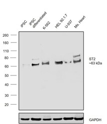
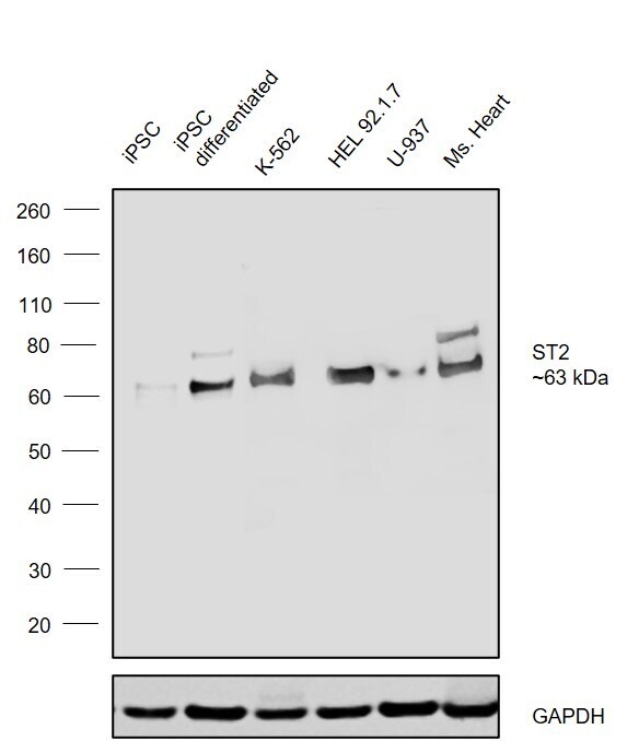
- Western blot was performed using Anti-ST2 Polyclonal Antibody (Product # PA5-23316) and a ~63 kDa band corresponding to Interleukin-1 receptor-like 1 was observed across cell lines and tissues tested . Membrane enriched extracts (50 µg lysate) of iPSC (Lane 1), iPSC differentiated into cardiomyocytes (Lane 2), K-562 (Lane 3), HEL 92.1.7 (Lane 4), U-937 (Lane 5), Mouse Heart (Lane 6) were electrophoresed using NuPAGE™ 4-12% Bis-Tris Protein Gel (Product # NP0321BOX). Resolved proteins were then transferred onto a nitrocellulose membrane (Product # IB23001) by iBlot® 2 Dry Blotting System (Product # IB21001). The blot was probed with the primary antibody (1 µg/mL) and detected by chemiluminescence with Goat anti-Rabbit IgG (H+L) Superclonal™ Recombinant Secondary Antibody, HRP (Product # A27036,1:20000) using the iBright FL 1000 (Product # A32752). Chemiluminescent detection was performed using SuperSignal™ West Pico PLUS Chemiluminescent Substrate (Product # 34580).Relative expression observed in K562 as highest expressing model versus in U-937 as a low expressing model.
- Submitted by
- Invitrogen Antibodies (provider)
- Main image
- Experimental details
- Western blot analysis of ST2 in human kidney lysate in the 1) absence and 2) presence of immunizing peptide, 3) mouse kidney and 4) rat kidney lysate. Samples were incubated in ST2 polyclonal antibody (Product # PA5-23316) followed by a goat anti-rabbit Ig HRP secondary antibody. PicoTect ECL substrate solution was used for this test.
Supportive validation
- Submitted by
- Invitrogen Antibodies (provider)
- Main image
- Experimental details
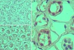
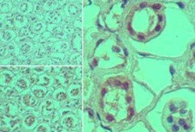
- Immunohistochemical analysis of ST2 in formalin-fixed, paraffin-embedded human kidney tissue. Samples were incubated in ST2 polyclonal antibody (Product # PA5-23316) using a dilution of 5 µg/mL. Isotype control (top left) and this antibody (bottom left, right).
Supportive validation
- Submitted by
- Invitrogen Antibodies (provider)
- Main image
- Experimental details
- NULL

- Submitted by
- Invitrogen Antibodies (provider)
- Main image
- Experimental details
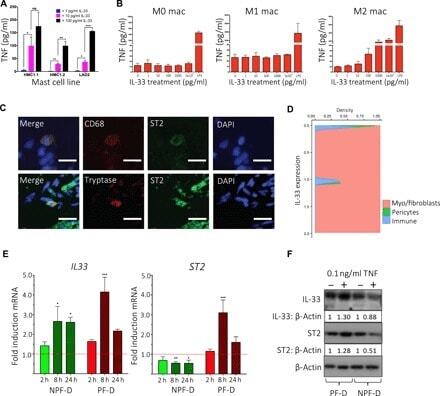
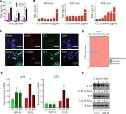
- Fig. 3 Stromal-immune cell crosstalk mediated by IL-33 and TNF. ( A ) Bar plot of enzyme-linked immunosorbent assay (ELISA) showing TNF secretion in mast cell lines (HMC1.1, HMC1.2, and LAD2) following stimulation with increasing doses of rIL-33 ( n = 3, mean +- SEM). ( B ) Bar plots of ELISA showing TNF secretion by human monocytes and macrophages following stimulation with increasing doses of rIL-33 ( n = 3 independent donors, mean +- SEM). ( C ) Representative confocal images of immunofluorescence for ST2 expression on mast cells (tryptase) and macrophages (CD68) in DD nodules. Scale bars, 10 mum. ( D ) Conditional density plot of single-cell RNA-seq dataset showing IL33 expression by labeled cell types. Density is Gaussian kernel density estimate ( n = 6 DD patients, k = 7332 cells). ( E ) Bar plots showing IL33 and ST2 gene expression by PF-D and NPF-D dermal fibroblasts from DD patients following stimulation with rTNF at 2, 8, and 24 hours ( n = 3 DD patients, mean +- SEM). ( F ) Western blot analysis showing IL-33 and ST2 protein expression by PF-D and nonpalmar NPF-D dermal fibroblasts from DD patients following stimulation with rTNF(0.1 ng/ml) for 24 hours. * P < 0.05, ** P < 0.01, *** P < 0.001. ns, not significant.