 Explore
 Validate
 Learn
LearnPA5-28383
antibody from Invitrogen Antibodies
Targeting: IL1RL1
DER4, FIT-1, IL33R, ST2, ST2L, ST2V, T1
 Western blot
Antibody data
- Antibody Data
- Antigen structure
- References [0]
- Comments [0]
- Validations
- Western blot [4]
Submit
Validation data
Reference
Comment
Report error
- Product number
- PA5-28383 - Provider product page

- Provider
- Invitrogen Antibodies
- Product name
- ST2 Polyclonal Antibody
- Antibody type
- Polyclonal
- Antigen
- Recombinant protein fragment
- Description
- Recommended positive controls: Raw264.7, Rat colon. Store product as a concentrated solution. Centrifuge briefly prior to opening the vial.
- Reactivity
- Human, Mouse, Rat
- Host
- Rabbit
- Isotype
- IgG
- Vial size
- 100 µL
- Concentration
- 1 mg/mL
- Storage
- Store at 4°C short term. For long term storage, store at -20°C, avoiding freeze/thaw cycles.
No comments: Submit comment
Supportive validation
- Submitted by
- Invitrogen Antibodies (provider)
- Main image
- Experimental details
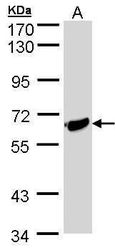
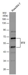
- Western blot analysis of ST2 using 30 µg of Raji lysate. Samples were loaded onto a 7.5% SDS-PAGE gel and probed with a ST2 polyclonal antibody (Product # PA5-28383) at a dilution of 1:500.
- Submitted by
- Invitrogen Antibodies (provider)
- Main image

- Experimental details
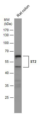
- Western Blot analysis of ST2 was performed by separating 50 µg of rat tissue extract by 10% SDS-PAGE. Proteins were transferred to a membrane and probed with a ST2 Polyclonal Antibody (Product # PA5-28383) at a dilution of 1:500.
- Submitted by
- Invitrogen Antibodies (provider)
- Main image
- Experimental details
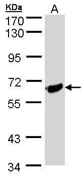
- Western Blot analysis of ST2 was performed by separating 30 µg of whole cell extract by 10% SDS-PAGE. Proteins were transferred to a membrane and probed with a ST2 Polyclonal Antibody (Product # PA5-28383) at a dilution of 1:500.

- Submitted by
- Invitrogen Antibodies (provider)
- Main image

- Experimental details
- Western blot was performed using Anti-ST2 Polyclonal Antibody (Product # PA5-28383) and a ~63kDa band corresponding to Interleukin-1 receptor-like 1 was observed across iPSCs. Membrane enriched extracts (50 µg lysate) of iPSC (Lane 1), iPSC differentiated to cardiomyocytes (Lane 2) were electrophoresed using NuPAGE™ 4-12% Bis-Tris Protein Gel (Product # NP0321BOX). Resolved proteins were then transferred onto a nitrocellulose membrane (Product # IB23001) by iBlot® 2 Dry Blotting System (Product # IB21001). The blot was probed with the primary antibody (1:1000) and detected by chemiluminescence with Goat anti-Rabbit IgG (H+L) Superclonal™ Recombinant Secondary Antibody, HRP (Product # A27036,1:20000) using the iBright FL 1000 (Product # A32752). Chemiluminescent detection was performed using SuperSignal™ West Pico PLUS Chemiluminescent Substrate (Product # 34580).Enhanced signal was observed in iPSCs differentiated into cardiomyocytes as compared to in undifferentiated iPSCs.
