 Explore
 Validate
 Learn
Learn Flow cytometry
Antibody data
- Antibody Data
- Antigen structure
- References [2]
- Comments [0]
- Validations
- Flow cytometry [1]
Submit
Validation data
Reference
Comment
Report error
- Product number
- FAB1216F-100 - Provider product page

- Provider
- R&D Systems
- Product name
- Mouse IL-18 R alpha/IL-1 R5 Fluorescein-conjugated Antibody
- Antibody type
- Monoclonal
- Description
- Protein A or G purified from hybridoma culture supernatant. Detects mouse IL-18 R alpha/IL-1 R5 in direct ELISAs and Western blots. In direct ELISAs and Western blots, no cross-reactivity with recombinant mouse (rm) IL-1 RI, rmIL-1 RII, recombinant human (rh) IL-1 R3, rhIL-1 R4, recombinant rat (rr) IL-1 R6, rmIL-1 R7, rhIL-1 R8, rhIL-1 R9, or rmSIGIRR is observed.
- Reactivity
- Mouse
- Host
- Rat
- Antigen sequence
Q61098- Isotype
- IgG
- Antibody clone number
- 112614
- Vial size
- 100 Tests
- Storage
- Protect from light. Do not freeze. 12 months from date of receipt, 2 to 8 °C as supplied.
Submitted references IL-18 acts in synergy with IL-7 to promote ex vivo expansion of T lymphoid progenitor cells.
Anti-inflammatory cytokines directly inhibit innate but not adaptive CD8+ T cell functions.
Gandhapudi SK, Tan C, Marino JH, Taylor AA, Pack CC, Gaikwad J, Van De Wiele CJ, Wren JD, Teague TK
Journal of immunology (Baltimore, Md. : 1950) 2015 Apr 15;194(8):3820-8
Journal of immunology (Baltimore, Md. : 1950) 2015 Apr 15;194(8):3820-8
Anti-inflammatory cytokines directly inhibit innate but not adaptive CD8+ T cell functions.
Freeman BE, Meyer C, Slifka MK
Journal of virology 2014 Jul;88(13):7474-84
Journal of virology 2014 Jul;88(13):7474-84
No comments: Submit comment
Supportive validation
- Submitted by
- R&D Systems (provider)
- Main image
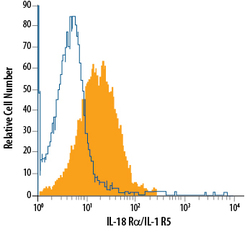
- Experimental details
- Detection of IL-18 R alpha/IL-1 R5 in Mouse Splenocytes by Flow Cytometry. Mouse splenocytes were stained with Rat Anti-Mouse IL-18 R alpha/IL-1 R5 Fluorescein-conjugated Monoclonal Antibody (Catalog # FAB1216F, filled histogram) or isotype control antibody (Catalog # IC005F, open histogram). View our protocol for Staining Membrane-associated Proteins.
