Explore
 Validate
 Learn
Learn Western blot
 ELISA
ELISAAntibody data
- Antibody Data
- Antigen structure
- References [1]
- Comments [0]
- Validations
- Western blot [1]
- Immunohistochemistry [1]
- Other assay [2]
Submit
Validation data
Reference
Comment
Report error
- Product number
- PA5-72875 - Provider product page

- Provider
- Invitrogen Antibodies
- Product name
- DEPDC1B Polyclonal Antibody
- Antibody type
- Polyclonal
- Antigen
- Synthetic peptide
- Reactivity
- Human
- Host
- Rabbit
- Isotype
- IgG
- Vial size
- 100 μg
- Concentration
- 1 mg/mL
- Storage
- Store at 4°C short term. For long term storage, store at -20°C, avoiding freeze/thaw cycles.
Submitted references DEPDC1B promotes migration and invasion in pancreatic ductal adenocarcinoma by activating the Akt/GSK3β/Snail pathway.
Liu X, Li T, Huang X, Wu W, Li J, Wei L, Qian Y, Xu H, Wang Q, Wang L
Oncology letters 2020 Nov;20(5):146
Oncology letters 2020 Nov;20(5):146
No comments: Submit comment
Supportive validation
- Submitted by
- Invitrogen Antibodies (provider)
- Main image

- Experimental details
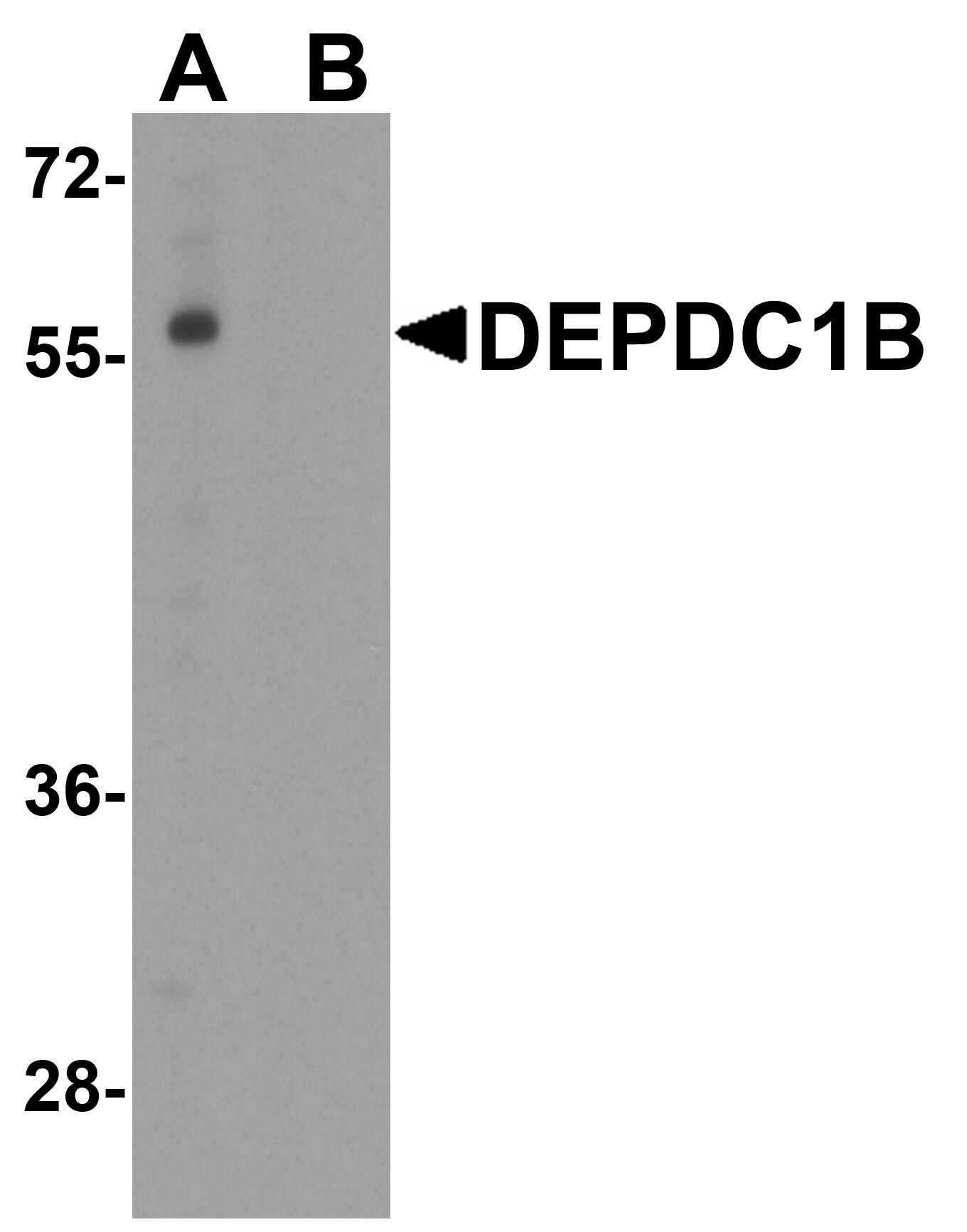
- Western Blot analysis of DEPDC1B in K562 cell lysate with DEPDC1B Polyclonal Antibody (Product # PA5-72875) at 1 µg/mL in (A) the absence and (B) the presence of blocking peptide.
Supportive validation
- Submitted by
- Invitrogen Antibodies (provider)
- Main image
- Experimental details
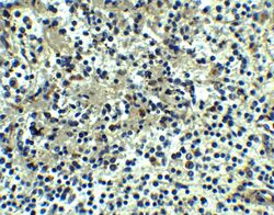
- Immunohistochemistry of DEPDC1B in human spleen tissue with DEPDC1B Polyclonal Antibody (Product # PA5-72875) at 5 µg/mL.

Supportive validation
- Submitted by
- Invitrogen Antibodies (provider)
- Main image

- Experimental details
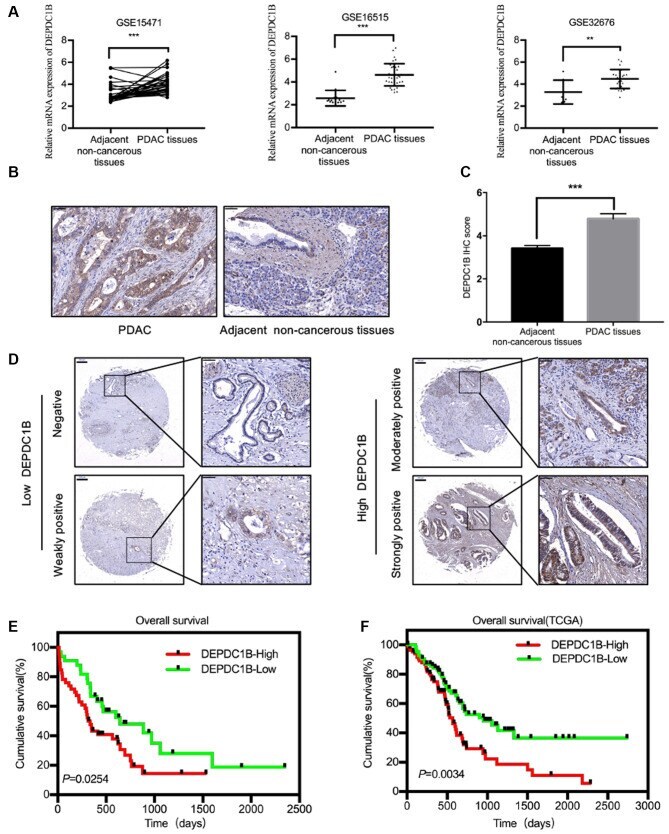
- Figure 1. DEPDC1B expression is increased in PDAC tissues. (A) The GEO datasets (GSE15471, GSE16515 and GSE32676) showed that the DEPDC1B mRNA expression levels were upregulated in PDAC tissues. (B) Representative IHC staining images for DEPDC1B in PDAC and adjacent non-cancerous tissues. Scale bar, 50 um. (C) DEPDC1B IHC score in PDAC tissues and adjacent non-cancerous tissues on TMAs. (D) Representative IHC staining images for DEPDC1B expression levels in TMAs. Scale bar, 200 um (left), 50 um (right). (E and F) Kaplan-Meier analysis showed the association between DEPDC1B expression and patient prognosis. **P
- Submitted by
- Invitrogen Antibodies (provider)
- Main image
- Experimental details
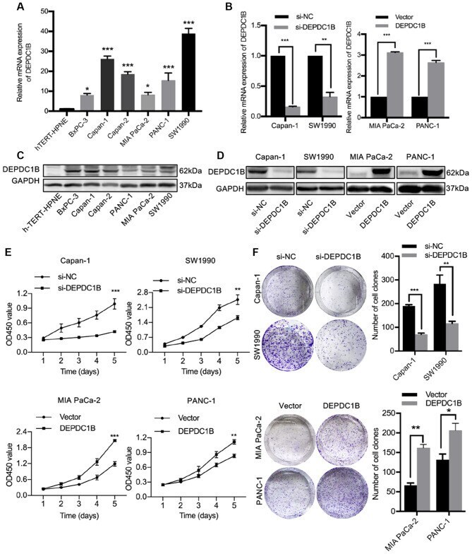
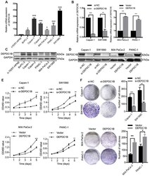
- Figure 2. DEPDC1B expression in PDAC cell lines and the effect of DEPDC1B on PDAC cell proliferation. (A) The DEPDC1B mRNA expression levels were detected in PDAC cell lines and compared with normal pancreatic cells hTERT-HPNE. (B) The DEPDC1B knockdown and overexpression efficiencies were measured by reverse transcription-quantitative PCR. (C) The DEPDC1B protein expression levels were detected in hTERT-HPNE and several PDAC cell lines by western blotting. (D) The DEPDC1B knockdown and overexpression efficiencies were measured by western blotting. (E) Cell Counting Kit-8 assays demonstrated that DEPDC1B knockdown suppressed PDAC cell proliferation, whereas the overexpression of DEPDC1B promoted PDAC cell proliferation. (F) DEPDC1B knockdown inhibited colony formation of PDAC cells, but the overexpression of DEPDC1B increased colony formation. *P