Explore
 Validate
 Learn
Learn Western blot
 Immunocytochemistry
Antibody data
- Antibody Data
- Antigen structure
- References [2]
- Comments [0]
- Validations
- Immunocytochemistry [1]
- Immunohistochemistry [1]
Submit
Validation data
Reference
Comment
Report error
- Product number
- HPA044682 - Provider product page

- Provider
- Atlas Antibodies
- Proper citation
- Atlas Antibodies Cat#HPA044682, RRID:AB_2679044
- Product name
- Anti-PLIN4
- Antibody type
- Polyclonal
- Description
- Polyclonal Antibody against Human PLIN4, Gene description: perilipin 4, Alternative Gene Names: KIAA1881, S3-12, Validated applications: ICC, IHC, WB, Uniprot ID: Q96Q06, Storage: Store at +4°C for short term storage. Long time storage is recommended at -20°C.
- Reactivity
- Human
- Host
- Rabbit
- Conjugate
- Unconjugated
- Isotype
- IgG
- Vial size
- 100 µl
- Concentration
- 0.1 mg/ml
- Storage
- Store at +4°C for short term storage. Long time storage is recommended at -20°C.
- Handling
- The antibody solution should be gently mixed before use.
Submitted references Senp7 deficiency impairs lipid droplets maturation in white adipose tissues via Plin4 deSUMOylation
Perilipin 4 in human skeletal muscle: localization and effect of physical activity
Pei J, Zou D, Li L, Kang L, Sun M, Li X, Chen Q, Chen D, Qu B, Gao X, Lin Z
Journal of Biological Chemistry 2024;300(6):107319
Journal of Biological Chemistry 2024;300(6):107319
Perilipin 4 in human skeletal muscle: localization and effect of physical activity
Pourteymour S, Lee S, Langleite T, Eckardt K, Hjorth M, Bindesbøll C, Dalen K, Birkeland K, Drevon C, Holen T, Norheim F
Physiological Reports 2015;3(8):e12481
Physiological Reports 2015;3(8):e12481
No comments: Submit comment
Supportive validation
- Submitted by
- Atlas Antibodies (provider)
- Main image

- Experimental details
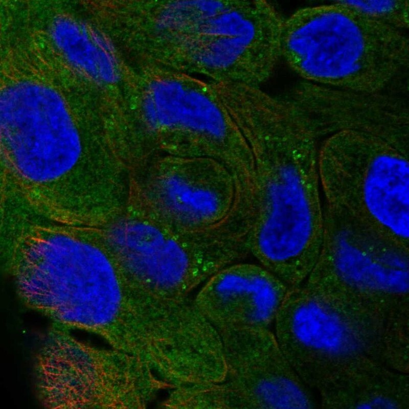
- Immunofluorescent staining of human cell line RT4 shows localization to plasma membrane, cytosol & lipid droplets.
- Sample type
- Human
Supportive validation
- Submitted by
- Atlas Antibodies (provider)
- Enhanced method
- Orthogonal validation
- Main image
- Experimental details
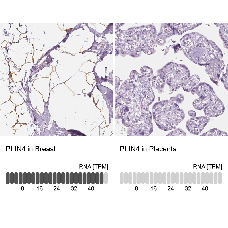
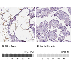
- Immunohistochemistry analysis in human breast and placenta tissues using HPA044682 antibody. Corresponding PLIN4 RNA-seq data are presented for the same tissues.
- Sample type
- Human
- Protocol
- Protocol