Explore
 Validate
 Learn
Learn Western blot
 Immunohistochemistry
Antibody data
- Antibody Data
- Antigen structure
- References [1]
- Comments [0]
- Validations
- Immunohistochemistry [1]
- Flow cytometry [2]
- Other assay [5]
Submit
Validation data
Reference
Comment
Report error
- Product number
- PA5-114352 - Provider product page

- Provider
- Invitrogen Antibodies
- Product name
- OXPAT Polyclonal Antibody
- Antibody type
- Polyclonal
- Antigen
- Recombinant full-length protein
- Reactivity
- Human, Mouse
- Host
- Rabbit
- Isotype
- IgG
- Vial size
- 100 μL
- Concentration
- 1 mg/mL
- Storage
- Store at 4°C short term. For long term storage, store at -20°C, avoiding freeze/thaw cycles.
Submitted references Leptin Reduces Plin5 m(6)A Methylation through FTO to Regulate Lipolysis in Piglets.
Wei D, Sun Q, Li Y, Li C, Li X, Sun C
International journal of molecular sciences 2021 Sep 30;22(19)
International journal of molecular sciences 2021 Sep 30;22(19)
No comments: Submit comment
Supportive validation
- Submitted by
- Invitrogen Antibodies (provider)
- Main image
- Experimental details
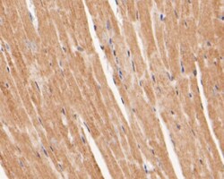
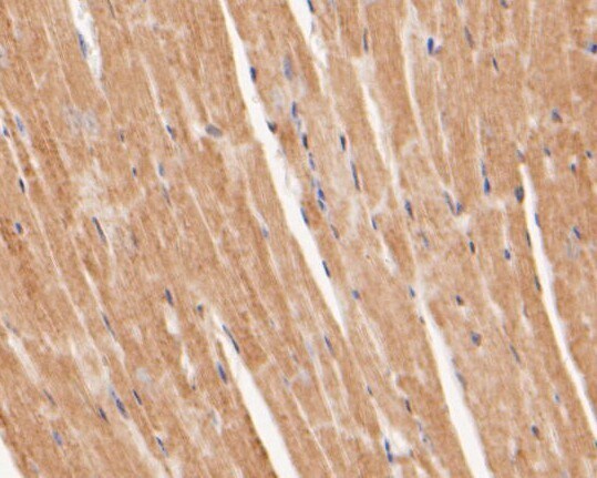
- Immunohistochemical analysis of OXPAT in paraffin-embedded mouse heart tissue using a polyclonal antibody (Product #PA5-114352). The section was pre-treated using heat mediated antigen retrieval with Tris-EDTA buffer (pH 8.0-8.4) for 20 minutes. The tissues were blocked in 5% BSA for 30 minutes at room temperature, washed with ddH2O and PBS, and then probed with the primary antibody (1:100) for 30 minutes at room temperature. The detection was performed using an HRP conjugated compact polymer system. DAB was used as the chromogen. Tissues were counterstained with hematoxylin and mounted with DPX.
Supportive validation
- Submitted by
- Invitrogen Antibodies (provider)
- Main image
- Experimental details
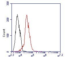
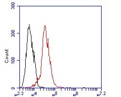
- Flow cytometric analysis of OXPAT using JAR cells and a OXPAT polyclonal antibody (Product #PA5-114352). The cells were fixed, permeabilized and stained with the primary antibody at a dilution of 1:100 (red). After incubation of the primary antibody at room temperature for an hour, the cells were stained with a Alexa Fluor 488-conjugated goat anti-rabbit IgG Secondary antibody at 1:500 dilution for 30 minutes. Unlabeled sample was used as a control (cells without incubation with primary antibody; black).

- Submitted by
- Invitrogen Antibodies (provider)
- Main image

- Experimental details
- Flow cytometric analysis of OXPAT using JAR cells and a OXPAT polyclonal antibody (Product #PA5-114352). The cells were fixed, permeabilized and stained with the primary antibody at a dilution of 1:100 (red). After incubation of the primary antibody at room temperature for an hour, the cells were stained with a Alexa Fluor 488-conjugated goat anti-rabbit IgG Secondary antibody at 1:500 dilution for 30 minutes. Unlabeled sample was used as a control (cells without incubation with primary antibody; black).
Supportive validation
- Submitted by
- Invitrogen Antibodies (provider)
- Main image
- Experimental details
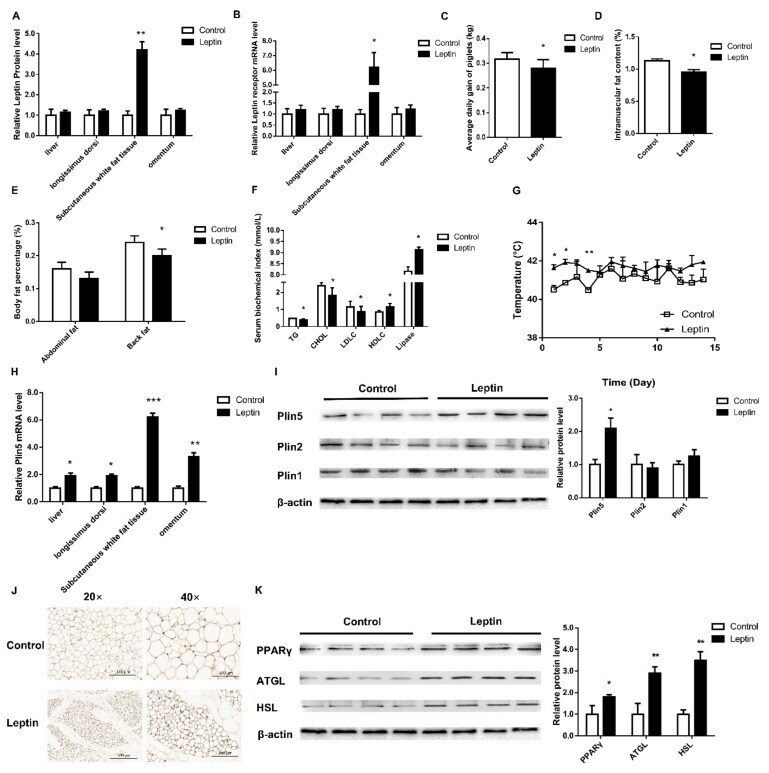
- Figure 1 Leptin improves the body fat composition and weight of piglets by up-regulating the expression of Plin5. ( A ) The leptin kit was used to detect the level of leptin protein in various tissues of piglets. ( B ) The expression of leptin receptors in various tissues of piglets. The effect of leptin treatment on: ( C ) the average daily gain, ( D ) the intramuscular fat content, ( E ) abdominal fat rate and back fat rate and ( F ) blood lipid level etc. ( G ) The change of piglet's body temperature for 15 days after leptin treatment. ( H , I ) The mRNA and protein expression levels of the Plin family were analyzed by real-time quantitative PCR and western blot. ( J ) Immunohistochemical detection of piglet adipose tissue. ( K ) Western blot analysis of lipolytic protein expression. n = 4 in each group, values are means +- SD. vs. control group, * p < 0.05, ** p < 0.01, *** p < 0.001.
- Submitted by
- Invitrogen Antibodies (provider)
- Main image
- Experimental details
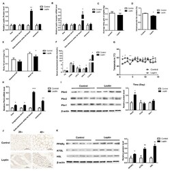
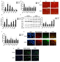
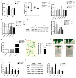
- Figure 2 Leptin promotes lipolysis by up-regulating the expression of Plin5 in pig adipocytes. ( A , B ) Real-time quantitative PCR was used to detect the mRNA expression of Plin5, lipolytic genes and mitochondrial complex-related genes in piglets subcutaneous adipose tissue. n = 4 in each group. ( C ) Oil red O staining was used to observe the number and size of LD in the subcutaneous fat tissue of piglets. ( D ) Real-time quantitative PCR was used to detect Plin5 and lipolytic gene expression in porcine adipocytes in vitro treated with 50 nmol/muL leptin for 24 h. The effect of 50 nmol/muL leptin treatment of porcine adipocytes in vitro on: ( E ) the expression of Plin5 and lipolytic protein and ( F ) the mRNA expression of mitochondrial complex-related genes. ( G , H ) Porcine adipocytes in vitro were treated with 50 nmol/muL leptin for 24 h to detect Plin5 immunofluorescence or BODIPY staining analysis. n = 3 for each group of cell samples, values are means +- SD. vs. control group, * p < 0.05, ** p < 0.01.

- Submitted by
- Invitrogen Antibodies (provider)
- Main image

- Experimental details
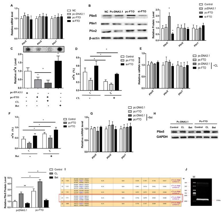
- Figure 4 FTO up-regulates Plin5 protein expression by inhibiting the m 6 A methylation of Plin5. The effect of FTO interference or overexpression in vitro on: ( A ) Plin family mRNA expression and ( B ) Plin family protein levels. ( C ) The relative M 6 A levels of pig adipocytes in vitro treated with CL and Bet after overexpression or interference with FTO. ( D ) The total M 6 A level of CL-treated pig adipocytes in vitro after overexpression or interference with FTO. ( E ) Plin family mRNA expression in CL-treated pig adipocytes in vitro after overexpression or interference with FTO. ( F ) The total M 6 A level of Bet-treated pig adipocytes after overexpression or interference with FTO. ( G ) Plin family mRNA expression in Bet-treated pig adipocytes in vitro after overexpression FTO. ( H ) Plin5 protein expression in pig adipocytes in vitro treated with CL and Bet after overexpression FTO. ( I , J ) Plin5 3'UTR end M 6 A site mutation vector construction. n = 4 for each group of cell samples, values are means +- SD. vs. control group, * p < 0.05, ** p < 0.01.
- Submitted by
- Invitrogen Antibodies (provider)
- Main image
- Experimental details
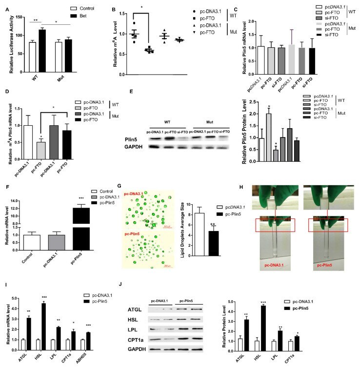
- Figure 5 Plin5 regulates lipid metabolism and energy metabolism through LD. ( A ) Luciferase reporter assay was used to analyze the effect of M 6 A mutation at the 3'UTR end of Plin5. ( B ) Relative M 6 A level analysis of wild-type and Plin5 mutant. ( C ) Detection of Plin5 mRNA expression of wild type and Plin5 mutant type. ( D ) Plin5 m 6 A methylation analysis of wild type and Plin5 mutant. ( E ) Plin5 protein expression of wild type and Plin5 mutant type. ( F ) Real-time quantitative PCR was used to detect the efficiency of the Plin5 overexpression vector. ( G ) Porcine adipocytes in vitro were transfected with Plin5 overexpression vector and then subjected to BODIPY fluorescent staining of LD. ( H ) Observation of lipid droplet extraction. ( I , J ) Porcine adipocytes in vitro were transfected with Plin5 over-expression vector to detect lipolysis-related genes mRNA or protein levels. n = 4 for each group of cell samples, values are means +- SD. vs. control group, * p < 0.05, ** p < 0.01, *** p < 0.001.
- Submitted by
- Invitrogen Antibodies (provider)
- Main image
- Experimental details
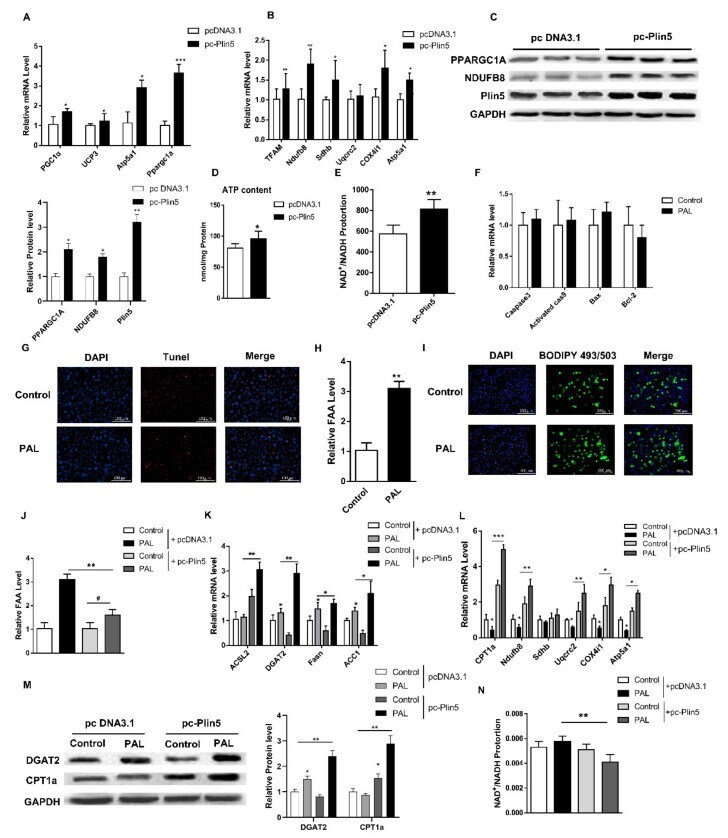
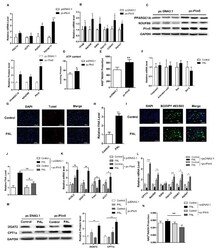
- Figure 6 Plin5 promotes lipid synthesis and enhances mitochondrial beta-oxidation in a lipotoxic model. ( A , B ) Real-time quantitative PCR was used to detect mitochondrial functional gene and beta-oxidation gene mRNA expression after Plin5 overexpression. ( C ) Under the same treatment as ( A , B ), the protein levels of differentially expressed genes were analyzed by Western blotting. ( D ) The kit detected the ATP content of porcine adipocytes transfected with Plin5 overexpression vector. ( E ) The ratio of NAD + /NADH was measured after Plin5 overexpression in porcine adipocytes in vitro. ( F ) 0.4 mmol/L PAL treated porcine adipocytes in vitro for 48 h to detect the mRNA expression of apoptosis-related genes. ( G ) Porcine adipocytes in vitro were treated with 0.4 mmol/L PAL for 48h to perform Tunel staining. ( H ) The FFA level was detected by the kit after treatment of 0.4 mmol/L PAL with porcine adipocytes in vitro for 48 h. ( I ) BODIPY staining after the same treatment in vitro. ( J ) After Plin5 overexpression, 0.4 mmol/L PAL was used to treat porcine adipocytes in vitro for 48 h to detect FFA levels. ( K , L ) After Plin5 was overexpressed, pig adipocytes in vitro were treated with 0.4 mmol/L PAL for 48 h to detect the mRNA levels of lipid synthesis genes and mitochondrial beta-oxidation genes. ( M ) Under the same treatment as ( K , L ), the protein levels of GDAT2 and CPT1a were analyzed by Western blotting. ( N ) The NAD + /NADH ratio is measured after the sam