Explore
 Validate
 Learn
Learn Western blot
 ELISA
ELISA Immunocytochemistry
Antibody data
- Antibody Data
- Antigen structure
- References [1]
- Comments [0]
- Validations
- Immunocytochemistry [2]
- Immunoprecipitation [1]
- Other assay [3]
Submit
Validation data
Reference
Comment
Report error
- Product number
- 41-3400 - Provider product page

- Provider
- Invitrogen Antibodies
- Product name
- Ninein Monoclonal Antibody (79.160-7)
- Antibody type
- Monoclonal
- Antigen
- Synthetic peptide
- Reactivity
- Human, Bovine
- Host
- Mouse
- Isotype
- IgG
- Antibody clone number
- 79.160-7
- Vial size
- 100 μg
- Concentration
- 0.5 mg/mL
- Storage
- -20°C
Submitted references NANOG/NANOGP8 Localizes at the Centrosome and is Spatiotemporally Associated with Centriole Maturation.
Mikulenkova E, Neradil J, Vymazal O, Skoda J, Veselska R
Cells 2020 Mar 11;9(3)
Cells 2020 Mar 11;9(3)
No comments: Submit comment
Supportive validation
- Submitted by
- Invitrogen Antibodies (provider)
- Main image
- Experimental details
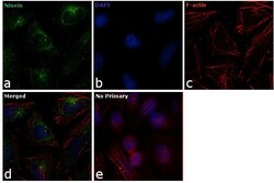
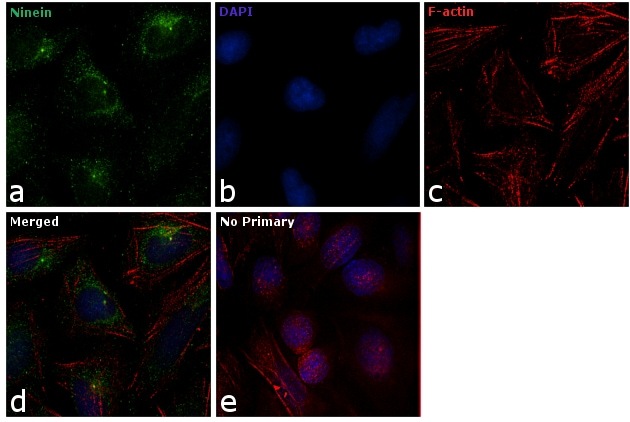
- Immunofluorescence analysis of Ninein was performed using 70% confluent log phase HeLa cells. The cells were fixed with 4% paraformaldehyde for 10 minutes, permeabilized with 0.1% Triton™ X-100 for 10 minutes, and blocked with 1% BSA for 1 hour at room temperature. The cells were labeled with Ninein Mouse Monoclonal Antibody (Product # 41-3400) at 5µg/mL in 0.1% BSA and incubated for 3 hours at room temperature and then labeled with Goat anti-Mouse IgG (H+L) Superclonal™ Secondary Antibody, Alexa Fluor® 488 conjugate (Product # A28175) a dilution of 1:2000 for 45 minutes at room temperature (Panel a: green). Nuclei (Panel b: blue) were stained with SlowFade® Gold Antifade Mountant with DAPI (Product # S36938). F-actin (Panel c: red) was stained with Alexa Fluor® 555 Rhodamine Phalloidin (Product # R415, 1:300). Panel d represents the merged image showing cytoplasm and centrosome localization. Panel e shows the no primary antibody control. The images were captured at 60X magnification.
- Submitted by
- Invitrogen Antibodies (provider)
- Main image

- Experimental details
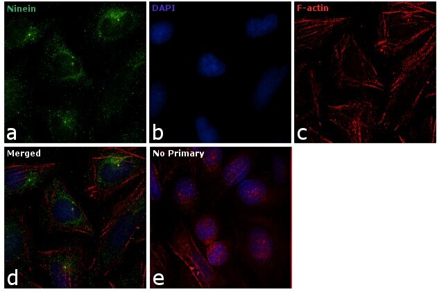
- Immunofluorescence analysis of Ninein was performed using 70% confluent log phase HeLa cells. The cells were fixed with 4% paraformaldehyde for 10 minutes, permeabilized with 0.1% Triton™ X-100 for 10 minutes, and blocked with 1% BSA for 1 hour at room temperature. The cells were labeled with Ninein Mouse Monoclonal Antibody (Product # 41-3400) at 5µg/mL in 0.1% BSA and incubated for 3 hours at room temperature and then labeled with Goat anti-Mouse IgG (H+L) Superclonal™ Secondary Antibody, Alexa Fluor® 488 conjugate (Product # A28175) a dilution of 1:2000 for 45 minutes at room temperature (Panel a: green). Nuclei (Panel b: blue) were stained with SlowFade® Gold Antifade Mountant with DAPI (Product # S36938). F-actin (Panel c: red) was stained with Alexa Fluor® 555 Rhodamine Phalloidin (Product # R415, 1:300). Panel d represents the merged image showing cytoplasm and centrosome localization. Panel e shows the no primary antibody control. The images were captured at 60X magnification.
Supportive validation
- Submitted by
- Invitrogen Antibodies (provider)
- Main image

- Experimental details
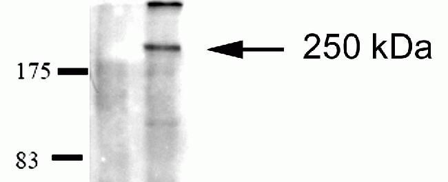
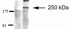
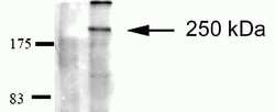
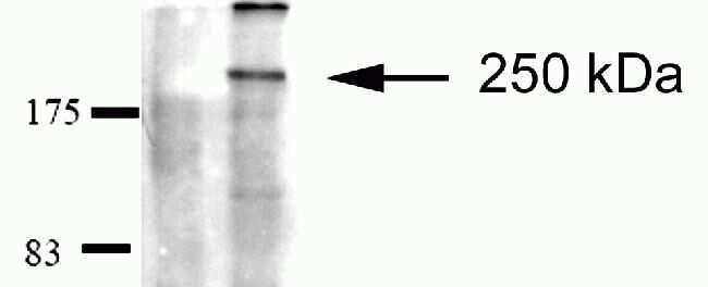
- Western blot analysis of HEK293 cell lysates after immunoprecipitaion (IP) using mouse anti-Ninein antibody (Product # 41-3400). Lane 1: Sample after IP using non-ninein related antibody. Lane 2: Sample after IP using ninein antibody.
Supportive validation
- Submitted by
- Invitrogen Antibodies (provider)
- Main image
- Experimental details
- Western blot analysis of HEK293 cell lysates after immunoprecipitaion (IP) using mouse anti-Ninein antibody (Product # 41-3400). Lane 1: Sample after IP using non-ninein related antibody. Lane 2: Sample after IP using ninein antibody.
- Submitted by
- Invitrogen Antibodies (provider)
- Main image

- Experimental details
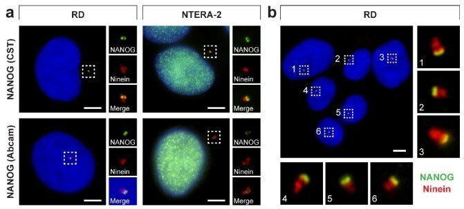
- Figure 9 NANOG colocalization with the mother centriole. ( a ) A colocalization of NANOG (green; #4903, Cell Signaling Technology (CST) and #ab109250, Abcam) with the mother centriole was detected using anti-ninein antibody (red), recognizing a protein of the mother centriole subdistal appendages. The nuclei were counterstained with Hoechst 33342 (blue). For each image, regions of interest and the respective close-ups are indicated by the dashed boxes. ( b ) NANOG signal (green; #4903, Cell Signaling Technology) partially colocalized with the ""double-spot"" signal of ninein (red). The nuclei were counterstained with Hoechst 33342 (blue). Respective close-ups of NANOG and ninein colocalization are indicated by numbers. Scale bars, 5 mum.