 Explore
 Validate
 Learn
Learn Western blot
Antibody data
- Antibody Data
- Antigen structure
- References [0]
- Comments [0]
- Validations
- Western blot [1]
- Immunocytochemistry [1]
- Immunohistochemistry [1]
Submit
Validation data
Reference
Comment
Report error
- Product number
- AP6329f - Provider product page

- Provider
- Abcepta
- Proper citation
- Abgent Cat#AP6329f, RRID:AB_2079406
- Product name
- CLC4 Antibody (C-term)
- Antibody type
- Polyclonal
- Antigen
- Synthetic peptide
- Description
- Purified Rabbit Polyclonal Antibody (Pab)
- Reactivity
- Human
- Host
- Rabbit
- Isotype
- IgG
- Vial size
- 400 µl
- Concentration
- 1.14 mg/ml
- Storage
- Maintain refrigerated at 2-8°C for up to 6 months. For long term storage store at -20°C in small aliquots to prevent freeze-thaw cycles.
No comments: Submit comment
Supportive validation
- Submitted by
- Abcepta (provider)
- Main image

- Experimental details
- "Western blot of chicken brain tissue incubated with CLC4 Antibody (C-term) (Cat.# AP6329f). Data courtesy of Emily McMains, Louisiana State University."
- Primary Ab dilution
- 1:1000

Supportive validation
- Submitted by
- Abcepta (provider)
- Main image
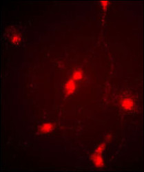
- Experimental details
- "Immunofluorescence image of cultured chick retinal amacrine (neuronal) cells labeled with CLC4 Antibody (C-term) (Cat # AP6329f). Data courtesy of Emily McMains, Louisiana State University."
- Primary Ab dilution
- 1:10~50

Supportive validation
- Submitted by
- Abcepta (provider)
- Main image

- Experimental details
- "Retinae were collected from adult White Leghorn chicken and fixed in the eyecup for two hours in 4% Paraformaldehyde (in PBS). Retinae were then removed from the eyecups and incubated in 30% sucrose (in PBS) overnight. Retinal tissue was embedded and frozen in OCT compound and cut into ~15um sections on a cryotome. Sections were blocked in 5% normal goat serum (in 1%BSA/.1%saponin PBS) for one hour and then incubated at RT with 1:250 (1%BSA/.1% saponin PBS) ClC4 antibody (Cat.# AP6329f) for 1 hour. Sections were then washed in PBS (3X10minutes) and then treated with secondary antibody (1:500 Cy3) for one hour. After another PBS wash series, sections were coverslipped and antibody labeling was visualized at 20X with a Leica upright microscope using a TRITC filter set and Xenon lamp illumination. (Crousillac et al, 2003)"
- Primary Ab dilution
- 1:10~50
