 Explore
 Validate
 Learn
Learn Western blot
 Immunocytochemistry
Antibody data
- Antibody Data
- Antigen structure
- References [1]
- Comments [0]
- Validations
- Immunocytochemistry [1]
- Immunohistochemistry [1]
Submit
Validation data
Reference
Comment
Report error
- Product number
- PA5-13348 - Provider product page

- Provider
- Invitrogen Antibodies
- Product name
- Anti-CLCN4 Polyclonal Antibody
- Antibody type
- Polyclonal
- Antigen
- Synthetic peptide
- Reactivity
- Human, Chicken/Avian
- Host
- Rabbit
- Isotype
- IgG
- Vial size
- 400 µL
- Concentration
- Lot Dependent
- Storage
- -20° C, Avoid Freeze/Thaw Cycles
Submitted references Prolactin stimulates sodium and chloride ion channels in A6 renal epithelial cells.
Greenlee MM, Mitzelfelt JD, Duke BJ, Al-Khalili O, Bao HF, Eaton DC
American journal of physiology. Renal physiology 2015 Apr 1;308(7):F697-705
American journal of physiology. Renal physiology 2015 Apr 1;308(7):F697-705
No comments: Submit comment
Supportive validation
- Submitted by
- Invitrogen Antibodies (provider)
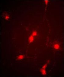
- Main image
- Experimental details
- Immunofluorescent analysis of cultured chick retinal amacrine (neuronal) cells labeled with anti-CLC4 polyclonal antibody (Product # PA5-13348).

Supportive validation
- Submitted by
- Invitrogen Antibodies (provider)
- Main image

- Experimental details
- Immunohistochemical analysis of CLC4 in retinae from adult White Leghorn chicken using a CLC4 polyclonal antibody (Product # PA5-13348). Retinae were fixed in the eyecup for two hours in 4% paraformaldehyde/PBS, then removed and incubated in 30% sucrose/PBS overnight. Retinal tissue was embedded and frozen in OCT compound and cut into ~15um sections on a cryotome. Sections were blocked in 5% normal goat serum (in 1% BSA/1% saponin PBS) for one hour and then incubated at room temperature with a CLC4 polyclonal antibody (Product # PA5-13348) at a dilution of 1:250. Sections were washed in PBS (3x10minutes) and then treated with secondary antibody (1:500 Cy3) for one hour, washed again and coverslipped. Antibody labeling was visualized at 20X with a Leica upright microscope using a TRITC filter set and Xenon lamp illumination.
