Explore
 Validate
 Learn
Learn Immunohistochemistry
Antibody data
- Antibody Data
- Antigen structure
- References [2]
- Comments [0]
- Validations
- Immunohistochemistry [1]
Submit
Validation data
Reference
Comment
Report error
- Product number
- HPA034525 - Provider product page

- Provider
- Atlas Antibodies
- Proper citation
- Atlas Antibodies Cat#HPA034525, RRID:AB_10669785
- Product name
- Anti-PTPRO
- Antibody type
- Polyclonal
- Description
- Polyclonal Antibody against Human PTPRO, Gene description: protein tyrosine phosphatase, receptor type, O, Alternative Gene Names: GLEPP1, NPHS6, PTP-oc, PTP-U2, PTPU2, Validated applications: IHC, Uniprot ID: Q16827, Storage: Store at +4°C for short term storage. Long time storage is recommended at -20°C.
- Reactivity
- Human
- Host
- Rabbit
- Conjugate
- Unconjugated
- Isotype
- IgG
- Vial size
- 100 µl
- Concentration
- 0.1 mg/ml
- Storage
- Store at +4°C for short term storage. Long time storage is recommended at -20°C.
- Handling
- The antibody solution should be gently mixed before use.
Submitted references Recommendations for mRNA analysis of micro-dissected glomerular tufts from paraffin-embedded human kidney biopsy samples
PTPRO-mediated autophagy prevents hepatosteatosis and tumorigenesis
Bockmeyer C, Wittig J, Säuberlich K, Selhausen P, Eßer M, Zeuschner P, Modde F, Amann K, Daniel C
BMC Molecular Biology 2018;19(1)
BMC Molecular Biology 2018;19(1)
PTPRO-mediated autophagy prevents hepatosteatosis and tumorigenesis
Zhang W, Hou J, Wang X, Jiang R, Yin Y, Ji J, Deng L, Huang X, Wang K, Sun B
Oncotarget 2015;6(11):9420-9433
Oncotarget 2015;6(11):9420-9433
No comments: Submit comment
Supportive validation
- Submitted by
- Atlas Antibodies (provider)
- Enhanced method
- Orthogonal validation
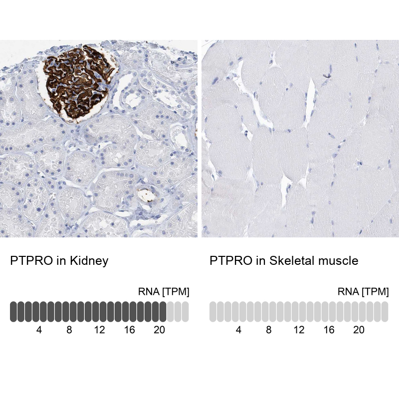
- Main image

- Experimental details
- Immunohistochemistry analysis in human kidney and skeletal muscle tissues using HPA034525 antibody. Corresponding PTPRO RNA-seq data are presented for the same tissues.
- Sample type
- Human
- Protocol
- Protocol