 Explore
 Validate
 Learn
Learn Western blot
 Immunohistochemistry
Antibody data
- Antibody Data
- Antigen structure
- References [5]
- Comments [0]
- Validations
- Western blot [1]
Submit
Validation data
Reference
Comment
Report error
- Product number
- HPA008546 - Provider product page

- Provider
- Atlas Antibodies
- Proper citation
- Atlas Antibodies Cat#HPA008546, RRID:AB_2066524
- Product name
- Anti-C2orf40
- Antibody type
- Polyclonal
- Description
- Polyclonal Antibody against Human C2orf40, Gene description: chromosome 2 open reading frame 40, Alternative Gene Names: augurin, ECRG4, Validated applications: WB, IHC, Uniprot ID: Q9H1Z8, Storage: Store at +4°C for short term storage. Long time storage is recommended at -20°C.
- Reactivity
- Human
- Host
- Rabbit
- Conjugate
- Unconjugated
- Isotype
- IgG
- Vial size
- 100 µl
- Concentration
- 0.1 mg/ml
- Storage
- Store at +4°C for short term storage. Long time storage is recommended at -20°C.
- Handling
- The antibody solution should be gently mixed before use.
Submitted references Loss of Ecrg4 improves calcium oxalate nephropathy
The ECRG4 cleavage product augurin binds the endotoxin receptor and influences the innate immune response during otitis media
ECRG4 expression in normal rat tissues: expression study and literature review
Thrombin-processed Ecrg4 recruits myeloid cells and induces antitumorigenic inflammation
Cell surface localization and release of the candidate tumor suppressor Ecrg4 from polymorphonuclear cells and monocytes activate macrophages
Theilig F, Cabuzu D, Ramakrishnan S, Moor M, Harmacek D, Auberson M, Durussel F, Bonny O
PLOS ONE 2022;17(10):e0275972
PLOS ONE 2022;17(10):e0275972
The ECRG4 cleavage product augurin binds the endotoxin receptor and influences the innate immune response during otitis media
Kurabi A, Hur D, Pak K, Gibson M, Webster N, Baird A, Eliceiri B, Ryan A
Frontiers in Genetics 2022;13
Frontiers in Genetics 2022;13
ECRG4 expression in normal rat tissues: expression study and literature review
Porzionato A, Rucinski M, Macchi V, Sarasin G, Malendowicz L, De Caro R
European Journal of Histochemistry 2015;59(2)
European Journal of Histochemistry 2015;59(2)
Thrombin-processed Ecrg4 recruits myeloid cells and induces antitumorigenic inflammation
Lee J, Dang X, Borboa A, Coimbra R, Baird A, Eliceiri B
Neuro-Oncology 2015;17(5):685-696
Neuro-Oncology 2015;17(5):685-696
Cell surface localization and release of the candidate tumor suppressor Ecrg4 from polymorphonuclear cells and monocytes activate macrophages
Baird A, Coimbra R, Dang X, Lopez N, Lee J, Krzyzaniak M, Winfield R, Potenza B, Eliceiri B
Journal of Leukocyte Biology 2012;91(5):773-781
Journal of Leukocyte Biology 2012;91(5):773-781
No comments: Submit comment
Enhanced validation
- Submitted by
- Atlas Antibodies (provider)
- Enhanced method
- Recombinant expression validation
- Main image
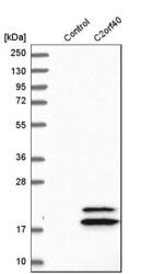
- Experimental details
- Western blot analysis in control (vector only transfected HEK293T lysate) and C2orf40 over-expression lysate (Co-expressed with a C-terminal myc-DDK tag (~3.1 kDa) in mammalian HEK293T cells, LY403168).
- Sample type
- Human
- Protocol
- Protocol
