Explore
 Validate
 Learn
Learn Western blot
Antibody data
- Antibody Data
- Antigen structure
- References [3]
- Comments [0]
- Validations
- Western blot [2]
- Immunocytochemistry [2]
- Immunohistochemistry [2]
Submit
Validation data
Reference
Comment
Report error
- Product number
- GTX109728 - Provider product page

- Provider
- GeneTex
- Proper citation
- GeneTex Cat#GTX109728, RRID:AB_1952096
- Product name
- SUCLA2 antibody [N2C3]
- Antibody type
- Polyclonal
- Reactivity
- Human, Mouse
- Host
- Rabbit
Submitted references Loss of succinyl-CoA synthase ADP-forming β subunit disrupts mtDNA stability and mitochondrial dynamics in neurons.
A Novel Role for Progesterone Receptor Membrane Component 1 (PGRMC1): A Partner and Regulator of Ferrochelatase.
Identification of the Mitochondrial Heme Metabolism Complex.
Zhao Y, Tian J, Sui S, Yuan X, Chen H, Qu C, Du Y, Guo L, Du H
Scientific reports 2017 Aug 2;7(1):7169
Scientific reports 2017 Aug 2;7(1):7169
A Novel Role for Progesterone Receptor Membrane Component 1 (PGRMC1): A Partner and Regulator of Ferrochelatase.
Piel RB 3rd, Shiferaw MT, Vashisht AA, Marcero JR, Praissman JL, Phillips JD, Wohlschlegel JA, Medlock AE
Biochemistry 2016 Sep 20;55(37):5204-17
Biochemistry 2016 Sep 20;55(37):5204-17
Identification of the Mitochondrial Heme Metabolism Complex.
Medlock AE, Shiferaw MT, Marcero JR, Vashisht AA, Wohlschlegel JA, Phillips JD, Dailey HA
PloS one 2015;10(8):e0135896
PloS one 2015;10(8):e0135896
No comments: Submit comment
Supportive validation
- Submitted by
- GeneTex (provider)
- Main image

- Experimental details
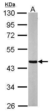
- Sample (20 ug of whole cell lysate) A: mouse brain 10% SDS PAGE GTX109728 diluted at 1:20000
- Submitted by
- GeneTex (provider)
- Main image
- Experimental details
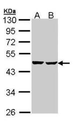
- Sample (30 ug of whole cell lysate) A: Hep G2 (GTX27900) B: Molt-4 (GTX27912) 10% SDS PAGE GTX109728 diluted at 1:1000
Supportive validation
- Submitted by
- GeneTex (provider)
- Main image
- Experimental details
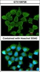
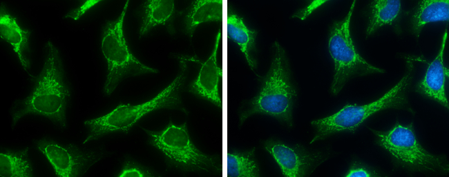
- Immunofluorescence analysis of methanol-fixed A431, using SUCLA2(GTX109728) antibody at 1:200 dilution.

- Submitted by
- GeneTex (provider)
- Main image

- Experimental details
- SUCLA2 antibody [N2C3] detects SUCLA2 protein at mitochondria by immunofluorescent analysis.Sample: HeLa cells were fixed in ice-cold MeOH for 5 min.Green: SUCLA2 protein stained by SUCLA2 antibody [N2C3] (GTX109728) diluted at 1:500.Blue: Hoechst 33342 staining.
Supportive validation
- Submitted by
- GeneTex (provider)
- Main image
- Experimental details
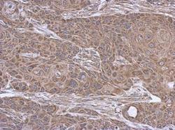
- Immunohistochemical analysis of paraffin-embedded Cal27 xenograft, using SUCLA2(GTX109728) antibody at 1:500 dilution.
- Submitted by
- GeneTex (provider)
- Main image
- Experimental details
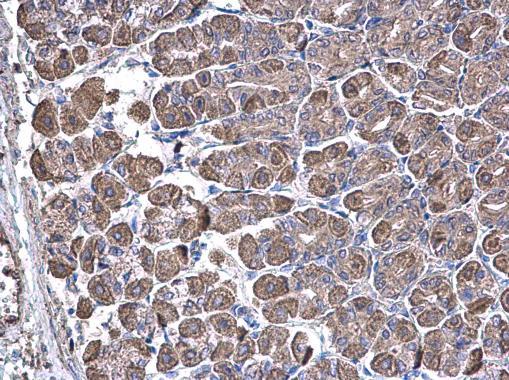
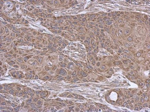
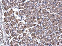
- SUCLA2 antibody [N2C3] detects SUCLA2 protein at cytoplasm on mouse stomach by immunohistochemical analysis. Sample: Paraffin-embedded mouse stomach. SUCLA2 antibody [N2C3] (GTX109728) diluted at 1:500.