Explore
 Validate
 Learn
Learn Western blot
Antibody data
- Antibody Data
- Antigen structure
- References [2]
- Comments [0]
- Validations
- Western blot [1]
- Immunocytochemistry [5]
- Immunohistochemistry [6]
- Other assay [1]
Submit
Validation data
Reference
Comment
Report error
- Product number
- PA5-21081 - Provider product page

- Provider
- Invitrogen Antibodies
- Product name
- TFF3 Polyclonal Antibody
- Antibody type
- Polyclonal
- Antigen
- Synthetic peptide
- Description
- A suggested positive control is rat small intestine tissue lysate. PA5-21081 can be used with blocking peptide PEP-1195.
- Reactivity
- Human, Mouse, Rat
- Host
- Rabbit
- Isotype
- IgG
- Vial size
- 100 μg
- Concentration
- 1 mg/mL
- Storage
- Maintain refrigerated at 2-8°C for up to 3 months. For long term storage store at -20°C
Submitted references Maternal administration of probiotics promotes gut development in mouse offsprings.
Conditioned medium from LS 174T goblet cells treated with oxyresveratrol strengthens tight junctions in Caco-2 cells.
Yu Y, Lu J, Oliphant K, Gupta N, Claud K, Lu L
PloS one 2020;15(8):e0237182
PloS one 2020;15(8):e0237182
Conditioned medium from LS 174T goblet cells treated with oxyresveratrol strengthens tight junctions in Caco-2 cells.
Hwang D, Jo H, Hwang S, Kim JK, Kim IH, Lim YH
Biomedicine & pharmacotherapy = Biomedecine & pharmacotherapie 2017 Jan;85:280-286
Biomedicine & pharmacotherapy = Biomedecine & pharmacotherapie 2017 Jan;85:280-286
No comments: Submit comment
Supportive validation
- Submitted by
- Invitrogen Antibodies (provider)
- Main image
- Experimental details
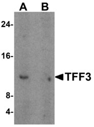
- Western Blot analysis of TFF3 in human colon tissue lysate with TFF3 Polyclonal Antibody (Product # PA5-21081) at 1 µg/mL in (A) the absence and (B) the presence of blocking peptide.
Supportive validation
- Submitted by
- Invitrogen Antibodies (provider)
- Main image
- Experimental details
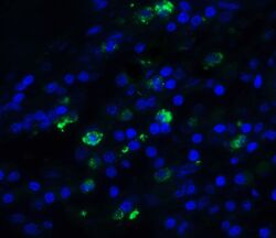
- Immunofluorescent analysis of human small intestine cells using a TFF3 polyclonal antibody (Product # PA5-21081) at a 20 µg/mL dilution.
- Submitted by
- Invitrogen Antibodies (provider)
- Main image

- Experimental details
- Immunocytochemistry of TFF3 in HeLa cells with TFF3 Polyclonal Antibody (Product # PA5-21081) at 2 µg/mL.
- Submitted by
- Invitrogen Antibodies (provider)
- Main image

- Experimental details
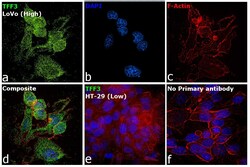
- Immunofluorescence analysis of Trefoil factor 3 was performed using 70% confluent log phase LoVo cells. The cells were fixed with 4% paraformaldehyde for 10 minutes, permeabilized with 0.1% Triton™ X-100 for 15 minutes, and blocked with 2% BSA for 45 minutes at room temperature. The cells were labeled with TFF3 Polyclonal Antibody (Product # PA5-21081) at 10 µg/mL in 0.1% BSA, incubated at 4-degree Celsius overnight, and then labeled with Donkey anti-Rabbit IgG (H+L) Highly Cross-Adsorbed Secondary Antibody, Alexa Fluor Plus 488 (Product # A32790), (1:2000 dilution), for 45 minutes at room temperature (Panel a: Green). Nuclei (Panel b: Blue) were stained with ProLong™ Diamond Antifade Mountant with DAPI (Product # P36962). F-actin (Panel c: Red) was stained with Rhodamine Phalloidin (Product # R415, 1:300 dilution). Panel d represents the merged image showing cytosolic localization. Panel e represents HT-29 cells showing no expression of TFF3. Panel f represents control cells with no primary antibody to assess the background. The images were captured at 60X magnification.

- Submitted by
- Invitrogen Antibodies (provider)
- Main image
- Experimental details
- Immunofluorescence analysis of Trefoil factor 3 was performed using 70% confluent log phase LoVo cells. The cells were fixed with 4% paraformaldehyde for 10 minutes, permeabilized with 0.1% Triton™ X-100 for 15 minutes, and blocked with 2% BSA for 45 minutes at room temperature. The cells were labeled with TFF3 Polyclonal Antibody (Product # PA5-21081) at 10 µg/mL in 0.1% BSA, incubated at 4-degree Celsius overnight, and then labeled with Donkey anti-Rabbit IgG (H+L) Highly Cross-Adsorbed Secondary Antibody, Alexa Fluor Plus 488 (Product # A32790), (1:2000 dilution), for 45 minutes at room temperature (Panel a: Green). Nuclei (Panel b: Blue) were stained with ProLong™ Diamond Antifade Mountant with DAPI (Product # P36962). F-actin (Panel c: Red) was stained with Rhodamine Phalloidin (Product # R415, 1:300 dilution). Panel d represents the merged image showing cytosolic localization. Panel e represents HT-29 cells showing no expression of TFF3. Panel f represents control cells with no primary antibody to assess the background. The images were captured at 60X magnification.

- Submitted by
- Invitrogen Antibodies (provider)
- Main image

- Experimental details
- Immunocytochemistry of TFF3 in HeLa cells with TFF3 Polyclonal Antibody (Product # PA5-21081) at 2 µg/mL.

Supportive validation
- Submitted by
- Invitrogen Antibodies (provider)
- Main image
- Experimental details
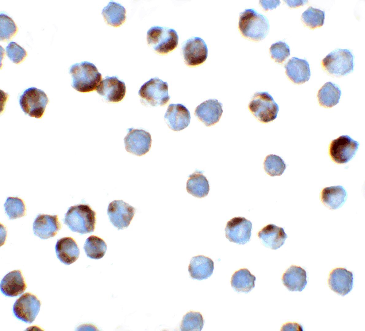
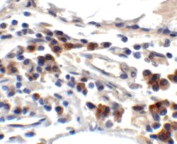
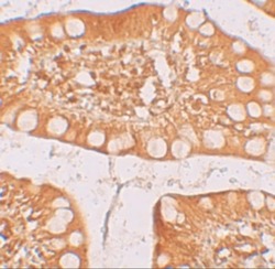
- Immunohistochemistry of TFF3 in human small intestine tissue with TFF3 Polyclonal Antibody (Product # PA5-21081) at 2 µg/mL.

- Submitted by
- Invitrogen Antibodies (provider)
- Main image

- Experimental details
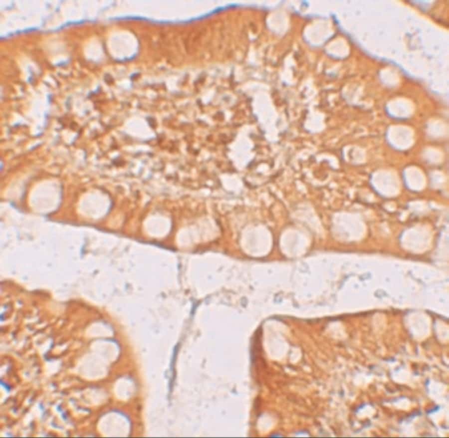
- Immunohistochemistry of TFF3 in human small intestine tissue with TFF3 Polyclonal Antibody (Product # PA5-21081) at 2.5 µg/mL.

- Submitted by
- Invitrogen Antibodies (provider)
- Main image
- Experimental details
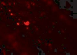
- Immunofluorescence of TFF3 in human small intestine tissue with TFF3 Polyclonal Antibody (Product # PA5-21081) at 20 µg/mL. Red: TFF3 Blue: DAPI staining

- Submitted by
- Invitrogen Antibodies (provider)
- Main image

- Experimental details
- Immunofluorescence of TFF3 in Human Small Intestine tissue with TFF3 Polyclonal Antibody (Product # PA5-21081) at 20 µg/mL.
- Submitted by
- Invitrogen Antibodies (provider)
- Main image
- Experimental details
- Immunohistochemistry of TFF3 in human small intestine tissue with TFF3 Polyclonal Antibody (Product # PA5-21081) at 2.5 µg/mL.
- Submitted by
- Invitrogen Antibodies (provider)
- Main image
- Experimental details
- Immunofluorescence of TFF3 in Human Small Intestine tissue with TFF3 Polyclonal Antibody (Product # PA5-21081) at 20 µg/mL.

Supportive validation
- Submitted by
- Invitrogen Antibodies (provider)
- Main image

- Experimental details
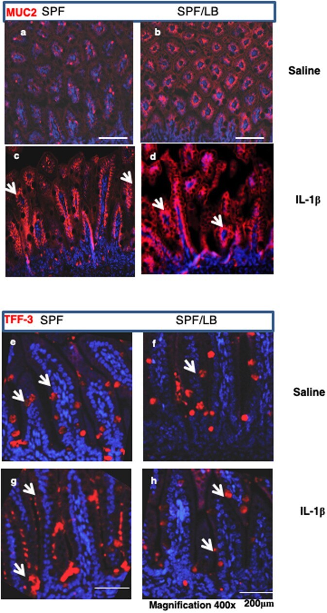
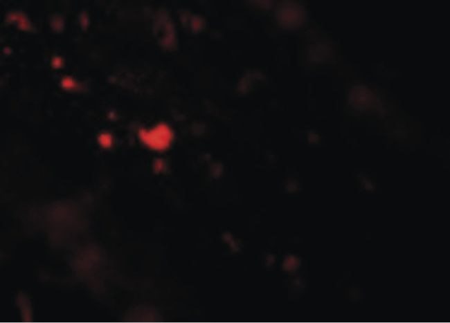
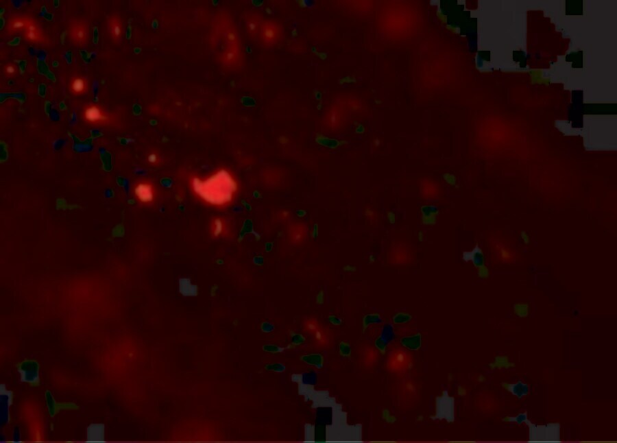
- 10.1371/journal.pone.0237182.g004 Fig 4 Maternal LB modulates IL-1beta-induced goblet cell Tff3 and MUC2 response to inflammatory stress in pre-weaned mice. a,b) Immunofluorescence detection of MUC2 molecules (red) in the mouse goblet cells in SPF (a) vs SPF/LB pups (b). Representative areas (n = 3 per group) are shown Bar 100 muM, magnification 400x. c,d) Representative areas of MUC2 exclusion from goblet cells in response to IL-1beta stimulation in SPF (c) or SPF/LB (d) pups are shown; magnification 400x. e,f) Representative area of immunofluorescence staining of TFF3 molecules (red) in the goblet cells. Left: SPF (e), and Right: SPF/LB (f) n = 3/per group) group, g,h) . IL-1beta induced TFF3 (red) secretion from goblet cells was detected by immunofluorescence staining. Representative areas were shown: Left: SPF ( g ), and Right: SPF/LB ( h ) (n = 3/per group) (magnification 400x).