Explore
 Validate
 Learn
Learn Western blot
 ELISA
ELISAAntibody data
- Antibody Data
- Antigen structure
- References [1]
- Comments [0]
- Validations
- Western blot [1]
- Immunocytochemistry [2]
- Immunohistochemistry [2]
- Other assay [1]
Submit
Validation data
Reference
Comment
Report error
- Product number
- PA5-98222 - Provider product page

- Provider
- Invitrogen Antibodies
- Product name
- TXK Polyclonal Antibody
- Antibody type
- Polyclonal
- Antigen
- Recombinant full-length protein
- Reactivity
- Human, Rat
- Host
- Rabbit
- Isotype
- IgG
- Vial size
- 100 μg
- Concentration
- 1 mg/mL
- Storage
- -20°C or -80°C if preferred
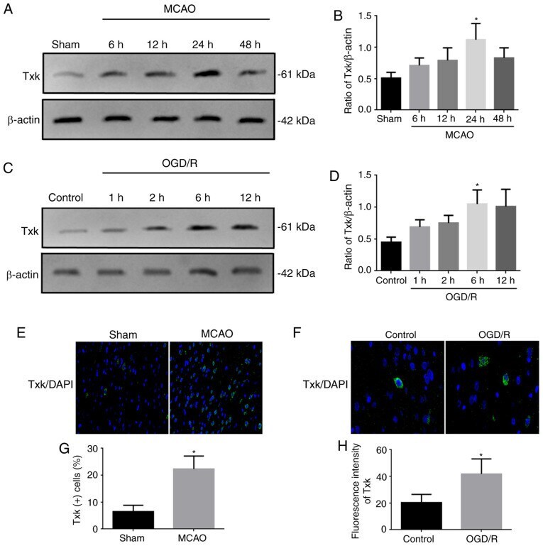
Submitted references Effects of Txk‑mediated activation of NF‑κB signaling pathway on neurological deficit and oxidative stress after ischemia‑reperfusion in rats.
Xu QL, Wu J
Molecular medicine reports 2021 Jul;24(1)
Molecular medicine reports 2021 Jul;24(1)
No comments: Submit comment
Supportive validation
- Submitted by
- Invitrogen Antibodies (provider)
- Main image

- Experimental details
- Western Blot analysis of TXK using a TXK Polyclonal antibody (Product # PA5-98222) at a concentration of 3 µg/mL. Positive WB detected in: HEK293 whole cell lysate, Jurkat whole cell lysate, Rat liver tissue, Rat kidney tissue. A secondary Goat polyclonal antibody to rabbit IgG was applied at a 1:50,000 dilution. Observed band size: 62 kDa.

Supportive validation
- Submitted by
- Invitrogen Antibodies (provider)
- Main image
- Experimental details
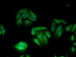
- Immunofluorescent analysis of TXK in HepG2 cells using a TXK polyclonal antibody (Product # PA5-98222) at a dilution of 1:100. Alexa Fluor 488-congugated Goat Anti-Rabbit IgG(H+L) secondary antibody was used.

- Submitted by
- Invitrogen Antibodies (provider)
- Main image

- Experimental details
- Immunofluorescent analysis of TXK in HepG2 cells using a TXK polyclonal antibody (Product # PA5-98222) at a dilution of 1:100. Alexa Fluor 488-congugated Goat Anti-Rabbit IgG(H+L) secondary antibody was used.

Supportive validation
- Submitted by
- Invitrogen Antibodies (provider)
- Main image
- Experimental details
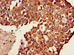
- Immunohistochemical analysis of TXK in paraffin embedded human breast cancer using a TXK polyclonal antibody (Product # PA5-98222) at a dilution of 1:100.

- Submitted by
- Invitrogen Antibodies (provider)
- Main image
- Experimental details
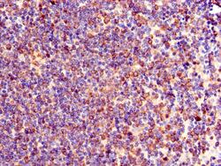
- Immunohistochemical analysis of TXK in paraffin embedded human lymph node tissue using a TXK polyclonal antibody (Product # PA5-98222) at a dilution of 1:100.

Supportive validation
- Submitted by
- Invitrogen Antibodies (provider)
- Main image
- Experimental details
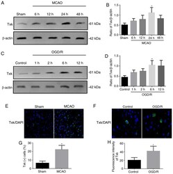
- Figure 1. Western blotting and immunofluorescence results. (A) Western blot analysis of Txk expression in sham and MCAO 6-, 12-, 24- and 48-h groups. (B) Quantification of Txk in different groups. (C) Western blot analysis of Txk expression in control and OGD/R 1-, 2-, 6- and 12-h neurons. (D) Quantification of Txk in different neurons. (E) Immunofluorescence assay of Txk in sham and MCAO rats. Magnification, x400) (F) Immunofluorescence assay of Txk in control and OGD/R neurons. Magnification, x1000 (G) Txk-positive cells in sham and MCAO rats and (H) fluorescence intensity in control and OGD/R neurons. Protein level was normalized to beta-actin. *P