Explore
 Validate
 Learn
Learn Western blot
Antibody data
- Antibody Data
- Antigen structure
- References [6]
- Comments [0]
- Validations
- Western blot [1]
- Immunohistochemistry [1]
Submit
Validation data
Reference
Comment
Report error
- Product number
- NB100-56355 - Provider product page

- Provider
- Novus Biologicals
- Proper citation
- Novus Cat#NB100-56355, RRID:AB_839113
- Product name
- Rabbit Polyclonal XAF1 Antibody
- Antibody type
- Polyclonal
- Description
- Protein G purified. In human liver, a 34 kDa band should be observed.
- Reactivity
- Human, Mouse
- Host
- Rabbit
- Isotype
- IgG
- Vial size
- 0.1 mg
- Concentration
- 0.5 mg/ml
- Storage
- Store at 4C short term. Aliquot and store at -20C long term. Avoid freeze-thaw cycles.
Submitted references SARM1 deficiency up-regulates XAF1, promotes neuronal apoptosis, and accelerates prion disease.
A co-clinical approach identifies mechanisms and potential therapies for androgen deprivation resistance in prostate cancer.
Down-regulation of the pro-apoptotic XIAP associated factor-1 (XAF1) during progression of clear-cell renal cancer.
XAF1 mediates tumor necrosis factor-alpha-induced apoptosis and X-linked inhibitor of apoptosis cleavage by acting through the mitochondrial pathway.
Xaf1 can cooperate with TNFalpha in the induction of apoptosis, independently of interaction with XIAP.
X-linked inhibitor of apoptosis (XIAP) protein protects against caspase activation and tissue loss after neonatal hypoxia-ischemia.
Zhu C, Li B, Frontzek K, Liu Y, Aguzzi A
The Journal of experimental medicine 2019 Apr 1;216(4):743-756
The Journal of experimental medicine 2019 Apr 1;216(4):743-756
A co-clinical approach identifies mechanisms and potential therapies for androgen deprivation resistance in prostate cancer.
Lunardi A, Ala U, Epping MT, Salmena L, Clohessy JG, Webster KA, Wang G, Mazzucchelli R, Bianconi M, Stack EC, Lis R, Patnaik A, Cantley LC, Bubley G, Cordon-Cardo C, Gerald WL, Montironi R, Signoretti S, Loda M, Nardella C, Pandolfi PP
Nature genetics 2013 Jul;45(7):747-55
Nature genetics 2013 Jul;45(7):747-55
Down-regulation of the pro-apoptotic XIAP associated factor-1 (XAF1) during progression of clear-cell renal cancer.
Kempkensteffen C, Fritzsche FR, Johannsen M, Weikert S, Hinz S, Dietel M, Riener MO, Moch H, Jung K, Krause H, Miller K, Kristiansen G
BMC cancer 2009 Aug 8;9:276
BMC cancer 2009 Aug 8;9:276
XAF1 mediates tumor necrosis factor-alpha-induced apoptosis and X-linked inhibitor of apoptosis cleavage by acting through the mitochondrial pathway.
Straszewski-Chavez SL, Visintin IP, Karassina N, Los G, Liston P, Halaban R, Fadiel A, Mor G
The Journal of biological chemistry 2007 Apr 27;282(17):13059-72
The Journal of biological chemistry 2007 Apr 27;282(17):13059-72
Xaf1 can cooperate with TNFalpha in the induction of apoptosis, independently of interaction with XIAP.
Xia Y, Novak R, Lewis J, Duckett CS, Phillips AC
Molecular and cellular biochemistry 2006 Jun;286(1-2):67-76
Molecular and cellular biochemistry 2006 Jun;286(1-2):67-76
X-linked inhibitor of apoptosis (XIAP) protein protects against caspase activation and tissue loss after neonatal hypoxia-ischemia.
Wang X, Zhu C, Wang X, Hagberg H, Korhonen L, Sandberg M, Lindholm D, Blomgren K
Neurobiology of disease 2004 Jun;16(1):179-89
Neurobiology of disease 2004 Jun;16(1):179-89
No comments: Submit comment
Supportive validation
- Submitted by
- Novus Biologicals (provider)
- Main image
- Experimental details
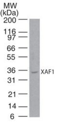
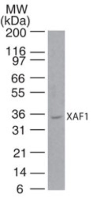
- Western Blot: XAF1 Antibody [NB100-56355] - analysis of XAF1 in human liver lysate using this antibody. I goat anti-rabbit Ig HRP secondary antibody and PicoTect ECL substrate solution were used for this test.
Supportive validation
- Submitted by
- Novus Biologicals (provider)
- Main image

- Experimental details
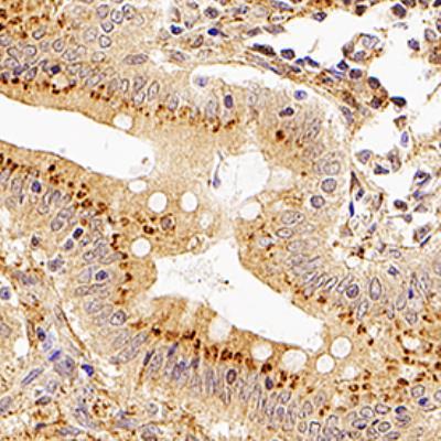
- Immunohistochemistry-Paraffin: XAF1 Antibody [NB100-56355] - XAF1 was detected in immersion fixed paraffin-embedded sections of human appendix using Rabbit Anti-Human XAF1 polyclonal Antibody (Catalog # NB100-56355) at 1:100 for 1 hour at room temperature followed by incubation with the Anti-Rabbit IgG VisUCyte™ HRP Polymer Antibody (Catalog # VC003). Tissue was stained using DAB (brown) and counterstained with hematoxylin (blue). Specific staining was localized to sperm cells.