Explore
 Validate
 Learn
Learn Western blot
Antibody data
- Antibody Data
- Antigen structure
- References [6]
- Comments [0]
- Validations
- Western blot [2]
- Immunocytochemistry [1]
Submit
Validation data
Reference
Comment
Report error
- Product number
- MA1-075 - Provider product page

- Provider
- Invitrogen Antibodies
- Product name
- LAP2 Monoclonal Antibody (RL29)
- Antibody type
- Monoclonal
- Antigen
- Purifed from natural sources
- Description
- MA1-075 has been successfully used in western blot and immunofluorescence applications to detect LAP2 from human and mouse samples. In western blot, this antibody predominantly detects the LAP2 delta subunit. The MA1-075 immunogen is full length rat LAP2, and has predicted reactivity with rat samples.
- Reactivity
- Human, Mouse, Rat
- Isotype
- IgG
- Antibody clone number
- RL29
- Vial size
- 100 µg
- Concentration
- 0.5 mg/mL
- Storage
- -20° C, Avoid Freeze/Thaw Cycles
Submitted references Lamin-binding fragment of LAP2 inhibits increase in nuclear volume during the cell cycle and progression into S phase.
Lamin-binding fragment of LAP2 inhibits increase in nuclear volume during the cell cycle and progression into S phase.
Integral membrane proteins of the nuclear envelope interact with lamins and chromosomes, and binding is modulated by mitotic phosphorylation.
Integral membrane proteins of the nuclear envelope interact with lamins and chromosomes, and binding is modulated by mitotic phosphorylation.
Integral membrane proteins specific to the inner nuclear membrane and associated with the nuclear lamina.
Integral membrane proteins specific to the inner nuclear membrane and associated with the nuclear lamina.
Yang L, Guan T, Gerace L
The Journal of cell biology 1997 Dec 1;139(5):1077-87
The Journal of cell biology 1997 Dec 1;139(5):1077-87
Lamin-binding fragment of LAP2 inhibits increase in nuclear volume during the cell cycle and progression into S phase.
Yang L, Guan T, Gerace L
The Journal of cell biology 1997 Dec 1;139(5):1077-87
The Journal of cell biology 1997 Dec 1;139(5):1077-87
Integral membrane proteins of the nuclear envelope interact with lamins and chromosomes, and binding is modulated by mitotic phosphorylation.
Foisner R, Gerace L
Cell 1993 Jul 2;73(7):1267-79
Cell 1993 Jul 2;73(7):1267-79
Integral membrane proteins of the nuclear envelope interact with lamins and chromosomes, and binding is modulated by mitotic phosphorylation.
Foisner R, Gerace L
Cell 1993 Jul 2;73(7):1267-79
Cell 1993 Jul 2;73(7):1267-79
Integral membrane proteins specific to the inner nuclear membrane and associated with the nuclear lamina.
Senior A, Gerace L
The Journal of cell biology 1988 Dec;107(6 Pt 1):2029-36
The Journal of cell biology 1988 Dec;107(6 Pt 1):2029-36
Integral membrane proteins specific to the inner nuclear membrane and associated with the nuclear lamina.
Senior A, Gerace L
The Journal of cell biology 1988 Dec;107(6 Pt 1):2029-36
The Journal of cell biology 1988 Dec;107(6 Pt 1):2029-36
No comments: Submit comment
Supportive validation
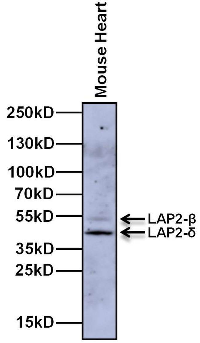
- Submitted by
- Invitrogen Antibodies (provider)
- Main image

- Experimental details
- Western blot analysis of LAP2 was performed by loading 20 µg of Mouse heart whole cell lysate and 10 µL of PageRuler Plus Prestained Protein Ladder (Product # 26619) onto a 4-20% Tris-Glycine polyacrylamide gel. Proteins were transferred to a nitrocellulose membrane and blocked with 5% Milk in TBST for at least 1 hour. LAP2-δ was detected at ~43 kDa and LAP2-β was detected at ~50 kDa using a LAP2 Armenian Hamster monoclonal antibody (Product # MA1-075) at a dilution of 4 µg/mL in 5% Milk in TBST overnight at 4C on a rocking platform, followed by a Goat anti-Armenian Hamster IgG (H+L) Secondary Antibody, HRP conjugate (Product # PA1-32045) at a dilution of 1:500 for at least 30 minutes at room temperature. Chemiluminescent detection was performed using SuperSignal West Dura substrate (Product # 34076) and the myECL Imager (Product # 62236).
- Submitted by
- Invitrogen Antibodies (provider)
- Main image

- Experimental details
- Western blot was performed using Anti-LAP2 Monoclonal Antibody (RL29) (Product # MA1-075) and a 27 kDa band corresponding to LAP2 was observed in Mouse Ovary, Mouse Heart, Rat Thymus, Rat Ovary and Rat Heart along with a uncharacterized band (*) at 50 kDa; and was absent in Mouse Brain and Rat Brain. Whole cell extracts (30 µg lysate) of Mouse Ovary (Lane 1), Mouse Heart (Lane 2), Mouse Brain (Lane 3), Rat Thymus (Lane 4), Rat Ovary (Lane 5), Rat Heart (Lane 6) and Rat Brain (Lane 7) were electrophoresed using NuPAGE™ 4-12% Bis-Tris Protein Gel (Product # NP0321BOX). Resolved proteins were then transferred onto a Nitrocellulose membrane (Product # LC2001) by iBlot® 2 Dry Blotting System (Product # IB21001). The blot was probed with the primary antibody (1:500 dilution) and detected by chemiluminescence with Rabbit anti-Hamster IgG (H+L Secondary Antibody, HRP (Product # A18889, 1:4000 dilution) using the iBright FL 1000 (Product # A32752). Chemiluminescent detection was performed using Novex® ECL Chemiluminescent Substrate Reagent Kit (Product # WP20005).

Supportive validation
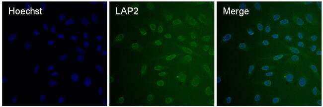
- Submitted by
- Invitrogen Antibodies (provider)
- Main image

- Experimental details
- Immunofluorescent analysis of LAP2 (green) in HeLa cells. The cells were fixed with 4% paraformaldehyde for 15 minutes, permeabilized with 0.1% Triton X-100 in PBS for 15 minutes, and blocked with 3% BSA in PBS (Product # 37525) for 30 minutes at room temperature. Cells were stained with a LAP2 Armenian Hamster monoclonal antibody (Product # MA1-075) at a dilution of 10 µg/mL in staining buffer for 1 hour at room temperature, and then incubated with a Goat anti-Armenian Hamster IgG Secondary Antibody, DyLight 488 conjugate at a dilution of 1:250 for 1 hour at room temperature (green). Nuclei (blue) were counterstained with Hoechst 33342 dye (Product # 62249). Images were taken on a Thermo Scientific ToxInsight Instrument at 20X magnification.