Explore
 Validate
 Learn
Learn Immunocytochemistry
 Immunohistochemistry
Antibody data
- Antibody Data
- Antigen structure
- References [1]
- Comments [0]
- Validations
- Immunocytochemistry [1]
Submit
Validation data
Reference
Comment
Report error
- Product number
- HPA036526 - Provider product page

- Provider
- Atlas Antibodies
- Proper citation
- Atlas Antibodies Cat#HPA036526, RRID:AB_10673499
- Product name
- Anti-SCFD2
- Antibody type
- Polyclonal
- Description
- Polyclonal Antibody against Human SCFD2, Gene description: sec1 family domain containing 2, Alternative Gene Names: FLJ39514, STXBP1L1, Validated applications: ICC, IHC, Uniprot ID: Q8WU76, Storage: Store at +4°C for short term storage. Long time storage is recommended at -20°C.
- Reactivity
- Human
- Host
- Rabbit
- Conjugate
- Unconjugated
- Isotype
- IgG
- Vial size
- 100 µl
- Concentration
- 0.2 mg/ml
- Storage
- Store at +4°C for short term storage. Long time storage is recommended at -20°C.
- Handling
- The antibody solution should be gently mixed before use.
Submitted references PSPC1 is a potential prognostic marker for hormone-dependent breast cancer patients and modulates RNA processing of ESR1 and SCFD2
Takeiwa T, Ikeda K, Suzuki T, Sato W, Iino K, Mitobe Y, Kawabata H, Horie K, Inoue S
Scientific Reports 2022;12(1)
Scientific Reports 2022;12(1)
No comments: Submit comment
Supportive validation
- Submitted by
- Atlas Antibodies (provider)
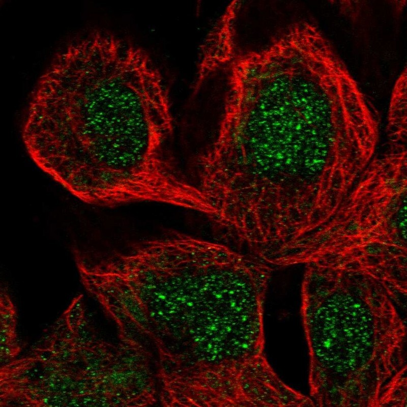
- Main image

- Experimental details
- Immunofluorescent staining of human cell line A-431 shows localization to nucleus.
- Sample type
- Human