Explore
 Validate
 Learn
Learn Western blot
Antibody data
- Antibody Data
- Antigen structure
- References [2]
- Comments [0]
- Validations
- Western blot [6]
- Immunoprecipitation [1]
- Immunohistochemistry [1]
Submit
Validation data
Reference
Comment
Report error
- Product number
- GTX101691 - Provider product page

- Provider
- GeneTex
- Proper citation
- GeneTex Cat#GTX101691, RRID:AB_1950586
- Product name
- ILK antibody [N1C1]
- Antibody type
- Polyclonal
- Reactivity
- Human, Mouse, Rat
- Host
- Rabbit
Submitted references Insulin-like growth factor-independent insulin-like growth factor binding protein 3 promotes cell migration and lymph node metastasis of oral squamous cell carcinoma cells by requirement of integrin β1.
Novel link of anti-apoptotic ATF3 with pro-apoptotic CTMP in the ischemic brain.
Yen YC, Hsiao JR, Jiang SS, Chang JS, Wang SH, Shen YY, Chen CH, Chang IS, Chang JY, Chen YW
Oncotarget 2015 Dec 8;6(39):41837-55
Oncotarget 2015 Dec 8;6(39):41837-55
Novel link of anti-apoptotic ATF3 with pro-apoptotic CTMP in the ischemic brain.
Huang CY, Chen JJ, Wu JS, Tsai HD, Lin H, Yan YT, Hsu CY, Ho YS, Lin TN
Molecular neurobiology 2015 Apr;51(2):543-57
Molecular neurobiology 2015 Apr;51(2):543-57
No comments: Submit comment
Supportive validation
- Submitted by
- GeneTex (provider)
- Main image
- Experimental details
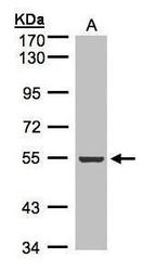
- Sample(30 £gg of whole cell lysate)A:A431(GTX27909) 7.5% SDS PAGEGTX101691 diluted at 1:500
- Submitted by
- GeneTex (provider)
- Main image
- Experimental details
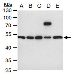
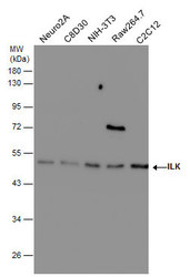
- ILK antibody [N1C1] detects ILK protein by western blot analysis.A. 30 £gg Neuro2A whole cell extract B. 30 £gg C8D30 whole cell extract C. 30 £gg NIH-3T3 whole cell extract D. 30 £gg Raw 264.7 whole cell extract E. 30 £gg C2Cl2 whole cell extract10 % SDS-PAGEILK antibody [N1C1] (GTX101691) dilution: 1:1000
- Submitted by
- GeneTex (provider)
- Main image

- Experimental details
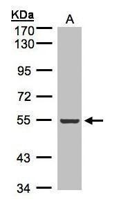
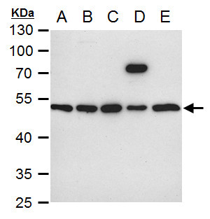
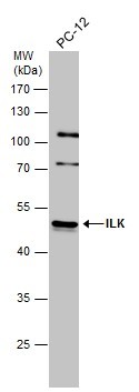
- Whole cell extract (30 £gg) was separated by 10% SDS-PAGE, and the membrane was blotted with ILK antibody [N1C1] (GTX101691) diluted at 1:500.
- Submitted by
- GeneTex (provider)
- Main image
- Experimental details
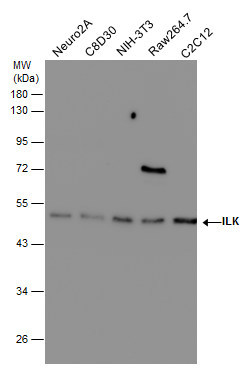
- Various whole cell extracts (30 £gg) were separated by 10% SDS-PAGE, and the membrane was blotted with ILK antibody [N1C1] (GTX101691) diluted at 1:500.
- Submitted by
- GeneTex (provider)
- Main image
- Experimental details
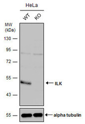
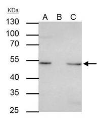
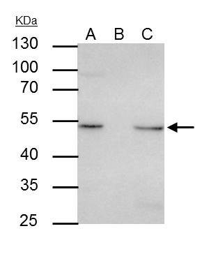
- Wild-type (WT) and ILK knockout (KO) HeLa cell extracts (30 ?g) were separated by 10% SDS-PAGE, and the membrane was blotted with ILK antibody [N1C1] (GTX101691) diluted at 1:500. The HRP-conjugated anti-rabbit IgG antibody (GTX213110-01) was used to detect the primary antibody.
- Submitted by
- GeneTex (provider)
- Main image
- Experimental details
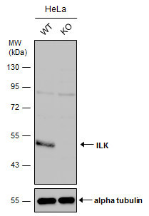
- Wild-type (WT) and ILK knockout (KO) HeLa cell extracts (30 ?g) were separated by 10% SDS-PAGE, and the membrane was blotted with ILK antibody [N1C1] (GTX101691) diluted at 1:500. The HRP-conjugated anti-rabbit IgG antibody (GTX213110-01) was used to detect the primary antibody.
Supportive validation
- Submitted by
- GeneTex (provider)
- Main image
- Experimental details
- ILK antibody [N1C1] immunoprecipitates ILK protein in IP experiments.IP samples: HeLa whole cell extractA. 30 £gg HeLa whole cell extractB. Control with 4 £gg of preimmune Rabbit IgGC. Immunoprecipitation of ILK protein by 4 £gg ILK antibody [N1C1] (GTX101691)10 % SDS-PAGEThe immunoprecipitated ILK protein was detected by ILK antibody [N1C1] (GTX101691) diluted at 1:500.[EasyBlot anti-rabbit IgG (GTX221666-01) was used as a secondary reagent]
Supportive validation
- Submitted by
- GeneTex (provider)
- Main image
- Experimental details
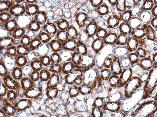
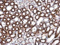
- ILK antibody [N1C1] detects ILK protein at cytoplasm in mouse kidney by immunohistochemical analysis. Sample: Paraffin-embedded mouse kidney. ILK antibody [N1C1] (GTX101691) diluted at 1:500.