 Explore
 Validate
 Learn
Learn Flow cytometry
Antibody data
- Antibody Data
- Antigen structure
- References [2]
- Comments [0]
- Validations
- Flow cytometry [2]
Submit
Validation data
Reference
Comment
Report error
- Product number
- 12-5838-42 - Provider product page

- Provider
- Invitrogen Antibodies
- Product name
- CD244 Monoclonal Antibody (eBioC1.7 (C1.7)), PE, eBioscience™
- Antibody type
- Monoclonal
- Antigen
- Other
- Description
- Description: The eBioC1.7 monoclonal antibody reacts with human CD244 (2B4, p38). In human, CD244 is a 38 kDa protein expressed on NK cells, a subset of CD8+ T cells, gammadelta T cells, monocytes, basophils and eosinophils. Binding of the CD244 ligand, CD48, results in NK cell activation, unlike mouse CD244, which is an inhibitory receptor. For CD244 expressed on NK cells, binding of CD48 results in enhanced NK cell cytotoxicity and secretion of IFN-gamma. Recently, it was demonstrated that binding of the C1.7 monoclonal antibody and CD48 involve the same residue in the V domain of human CD244, which explains the ability of C1.7 binding to induce activation of NK cells. Binding of C1.7 to CD244 leads to tyrosine phosphorylation and recruitment of the adaptor molecule SAP (SLAM-associated protein). Patients with X-linked lymphoproliferative disorder (XLPD) have a mutation in SAP which renders it unable to bind to phosphorylated CD244. Applications Reported: This eBioC1.7 (C1.7) antibody has been reported for use in flow cytometric analysis. Applications Tested: This eBioC1.7 (C1.7) antibody has been pre-titrated and tested by flow cytometric analysis of normal human peripheral blood cells. This can be used at 5 µL (0.125 µg) per test. A test is defined as the amount (µg) of antibody that will stain a cell sample in a final volume of 100 µL. Cell number should be determined empirically but can range from 10^5 to 10^8 cells/test. Excitation: 488-561 nm; Emission: 578 nm; Laser: Blue Laser, Green Laser, Yellow-Green Laser. Filtration: 0.2 µm post-manufacturing filtered.
- Reactivity
- Human
- Host
- Mouse
- Conjugate
- Yellow dye
- Isotype
- IgG
- Antibody clone number
- eBioC1.7 (C1.7)
- Vial size
- 100 Tests
- Concentration
- 5 µL/Test
- Storage
- 4° C, store in dark, DO NOT FREEZE!
Submitted references Exhaustion of the CD8(+) T Cell Compartment in Patients with Mutations in Phosphoinositide 3-Kinase Delta.
Human MAIT cells are xenobiotic-resistant, tissue-targeted, CD161hi IL-17-secreting T cells.
Wentink MWJ, Mueller YM, Dalm VASH, Driessen GJ, van Hagen PM, van Montfrans JM, van der Burg M, Katsikis PD
Frontiers in immunology 2018;9:446
Frontiers in immunology 2018;9:446
Human MAIT cells are xenobiotic-resistant, tissue-targeted, CD161hi IL-17-secreting T cells.
Dusseaux M, Martin E, Serriari N, Péguillet I, Premel V, Louis D, Milder M, Le Bourhis L, Soudais C, Treiner E, Lantz O
Blood 2011 Jan 27;117(4):1250-9
Blood 2011 Jan 27;117(4):1250-9
No comments: Submit comment
Supportive validation
- Submitted by
- Invitrogen Antibodies (provider)
- Main image
- Experimental details
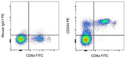
- Staining of normal human peripheral blood cells with Anti-Human CD8a FITC (Product # 11-0086-42) and Mouse IgG1 kappa Isotype Control PE (Product # 12-4714-81) (left) or Anti-Human CD244 PE (right). Cells in the lymphocyte gate were used for analysis.
- Conjugate
- Yellow dye

- Submitted by
- Invitrogen Antibodies (provider)
- Main image
- Experimental details
- Normal human peripheral blood cells were stained with CD8a Monoclonal Antibody, FITC (Product # 11-0087-42) and Mouse IgG1 kappa Isotype Control, PE (Product # 12-4714-82) (left) or CD244 Monoclonal Antibody, PE (right). Cells in the lymphocyte gate were used for analysis.
- Conjugate
- Yellow dye
