Explore
 Validate
 Learn
Learn Flow cytometry
Antibody data
- Antibody Data
- Antigen structure
- References [2]
- Comments [0]
- Validations
- Flow cytometry [1]
Submit
Validation data
Reference
Comment
Report error
- Product number
- 329508 - Provider product page

- Provider
- BioLegend
- Proper citation
- BioLegend Cat#329508, RRID:AB_1279190
- Product name
- PE anti-human CD244 (2B4)
- Antibody type
- Monoclonal
- Antigen
- CD244 (2B4)
- Description
- CD244, known as 2B4, is a 38 kD type I transmembrane protein. It is a member of the CD2 subset of the immunoglobulin superfamily (IgSF) molecules. CD244 is expressed on NK cells, a subset of T cells (including majority of CD8+ T cells and γ/δ T cells), monocytes, basophils, and eosinophils. CD48 is the ligand of CD244. It has been reported that ligation of human CD244 results in enhanced NK cell cytotoxicity and cytokine production. Recent studies have shown that human CD244, like murine CD244, has both activating and inhibitory functions, which are dependent on the density of surface 2B4 expression, degree of ligation, and the level of the adaptor molecule SAP expression.
- Reactivity
- Human
- Host
- Mouse
- Conjugate
- Yellow dye
- Isotype
- IgG
- Vial size
- 100 tests
- Storage
- The antibody solution should be stored undiluted at 4°C and protected from prolonged exposure to light. Do not freeze.
Submitted references Histone deacetylase inhibitors impair NK cell viability and effector functions through inhibition of activation and receptor expression.
2B4 engagement mediates rapid LFA-1 and actin-dependent NK cell adhesion to tumor cells as measured by single cell force spectroscopy.
Rossi LE, Avila DE, Spallanzani RG, Ziblat A, Fuertes MB, Lapyckyj L, Croci DO, Rabinovich GA, Domaica CI, Zwirner NW
Journal of leukocyte biology 2012 Feb;91(2):321-31
Journal of leukocyte biology 2012 Feb;91(2):321-31
2B4 engagement mediates rapid LFA-1 and actin-dependent NK cell adhesion to tumor cells as measured by single cell force spectroscopy.
Hoffmann SC, Cohnen A, Ludwig T, Watzl C
Journal of immunology (Baltimore, Md. : 1950) 2011 Mar 1;186(5):2757-64
Journal of immunology (Baltimore, Md. : 1950) 2011 Mar 1;186(5):2757-64
No comments: Submit comment
Supportive validation
- Submitted by
- BioLegend (provider)
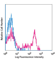
- Main image
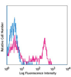
- Experimental details
- Human peripheral blood lymphocytes stained with C1.7 PE
- Conjugate
- Yellow dye