 Explore
 Validate
 Learn
Learn Western blot
 Immunocytochemistry
Antibody data
- Antibody Data
- Antigen structure
- References [2]
- Comments [0]
- Validations
- Immunocytochemistry [1]
Submit
Validation data
Reference
Comment
Report error
- Product number
- HPA002954 - Provider product page

- Provider
- Atlas Antibodies
- Proper citation
- Atlas Antibodies Cat#HPA002954, RRID:AB_1078997
- Product name
- Anti-GMFB
- Antibody type
- Polyclonal
- Description
- Polyclonal Antibody against Human GMFB, Gene description: glia maturation factor, beta, Alternative Gene Names: GMF, Validated applications: ICC, WB, IHC, Uniprot ID: P60983, Storage: Store at +4°C for short term storage. Long time storage is recommended at -20°C.
- Reactivity
- Human, Mouse, Rat
- Host
- Rabbit
- Conjugate
- Unconjugated
- Isotype
- IgG
- Vial size
- 100 µl
- Concentration
- 0.05 mg/ml
- Storage
- Store at +4°C for short term storage. Long time storage is recommended at -20°C.
- Handling
- The antibody solution should be gently mixed before use.
Submitted references GMFβ controls branched actin content and lamellipodial retraction in fibroblasts
Evaluation of Blastomere Biopsy Using a Mouse Model Indicates the Potential High Risk of Neurodegenerative Disorders in the Offspring
Haynes E, Asokan S, King S, Johnson H, Haugh J, Bear J
Journal of Cell Biology 2015;209(6):803-812
Journal of Cell Biology 2015;209(6):803-812
Evaluation of Blastomere Biopsy Using a Mouse Model Indicates the Potential High Risk of Neurodegenerative Disorders in the Offspring
Yu Y, Wu J, Fan Y, Lv Z, Guo X, Zhao C, Zhou R, Zhang Z, Wang F, Xiao M, Chen L, Zhu H, Chen W, Lin M, Liu J, Zhou Z, Wang L, Huo R, Zhou Q, Sha J
Molecular & Cellular Proteomics 2009;8(7):1490-1500
Molecular & Cellular Proteomics 2009;8(7):1490-1500
No comments: Submit comment
Supportive validation
- Submitted by
- Atlas Antibodies (provider)
- Main image
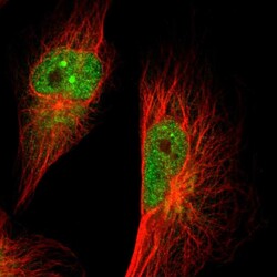
- Experimental details
- Immunofluorescent staining of human cell line U-251 MG shows localization to nucleoplasm, nuclear bodies & cytosol.
- Sample type
- Human
