Explore
 Validate
 Learn
Learn Immunocytochemistry
Antibody data
- Antibody Data
- Antigen structure
- References [1]
- Comments [0]
- Validations
- Immunocytochemistry [1]
- Immunohistochemistry [4]
- Other assay [1]
Submit
Validation data
Reference
Comment
Report error
- Product number
- PA5-61409 - Provider product page

- Provider
- Invitrogen Antibodies
- Product name
- TRMT6 Polyclonal Antibody
- Antibody type
- Polyclonal
- Antigen
- Recombinant protein fragment
- Description
- Immunogen sequence: KMIVMETCAG LVLGAMMERM GGFGSIIQLY PGGGPVRAAT ACFGFPKSFL SGLYEFPLNK VDSLLHGTFS AKMLSSEPKD SALVEESNGT LE Highest antigen sequence identity to the following orthologs: Mouse - 91%, Rat - 89%.
- Reactivity
- Human
- Host
- Rabbit
- Isotype
- IgG
- Vial size
- 100 μL
- Concentration
- 0.40 mg/mL
- Storage
- Store at 4°C short term. For long term storage, store at -20°C, avoiding freeze/thaw cycles.
Submitted references N(1)-methyladenosine methylation in tRNA drives liver tumourigenesis by regulating cholesterol metabolism.
Wang Y, Wang J, Li X, Xiong X, Wang J, Zhou Z, Zhu X, Gu Y, Dominissini D, He L, Tian Y, Yi C, Fan Z
Nature communications 2021 Nov 2;12(1):6314
Nature communications 2021 Nov 2;12(1):6314
No comments: Submit comment
Supportive validation
- Submitted by
- Invitrogen Antibodies (provider)
- Main image
- Experimental details
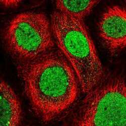
- Immunofluorecent analysis of TRMT6 in human cell line A-431 using TRMT6 Polyclonal Antibody (Product # PA5-61409). Staining shows localization to nucleus.

Supportive validation
- Submitted by
- Invitrogen Antibodies (provider)
- Main image
- Experimental details
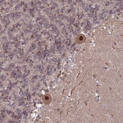
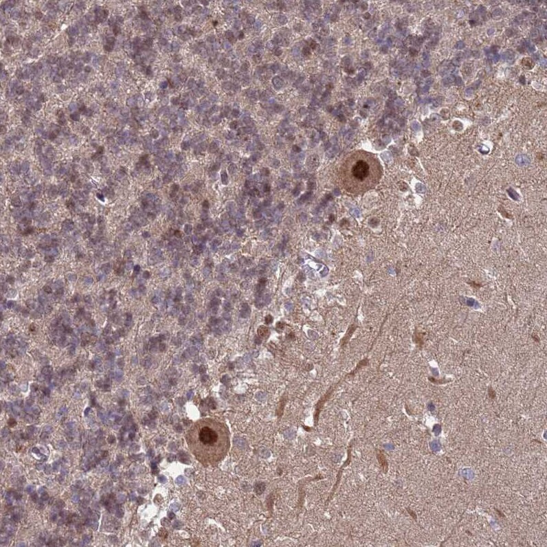
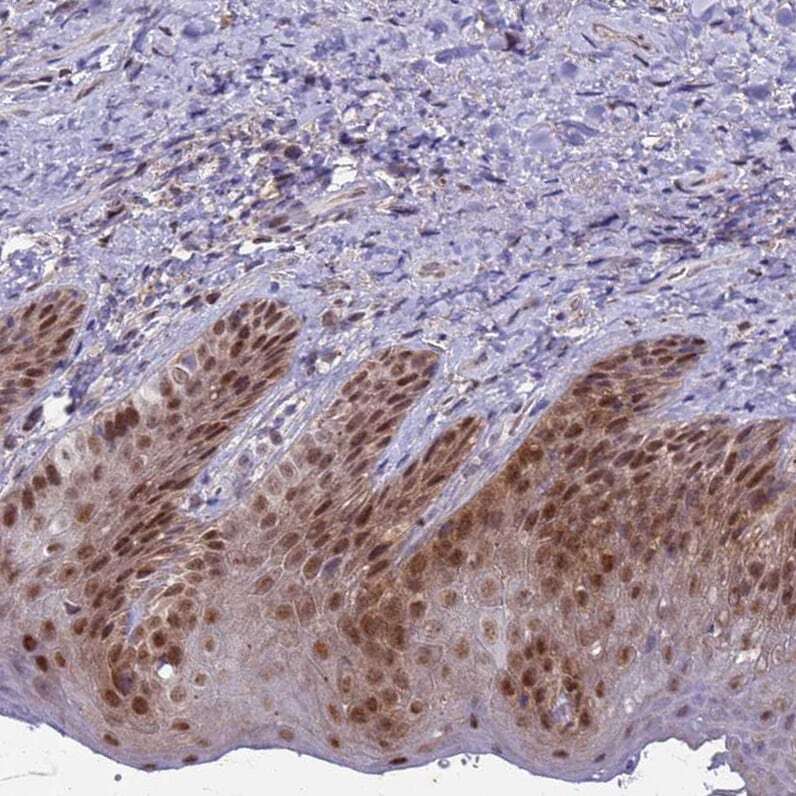
- Immunohistochemical analysis of TRMT6 in human cerebellum using TRMT6 Polyclonal Antibody (Product # PA5-61409) shows strong nuclear positivity in Purkinje cells.
- Submitted by
- Invitrogen Antibodies (provider)
- Main image

- Experimental details
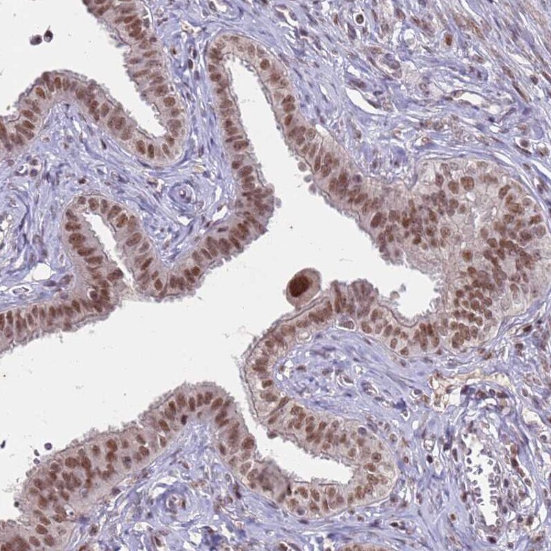
- Immunohistochemical analysis of TRMT6 in human fallopian tube using TRMT6 Polyclonal Antibody (Product # PA5-61409) shows moderate nuclear positivity in glandular cells.
- Submitted by
- Invitrogen Antibodies (provider)
- Main image
- Experimental details
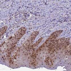
- Immunohistochemical analysis of TRMT6 in human skin using TRMT6 Polyclonal Antibody (Product # PA5-61409) shows strong nuclear positivity in epidermal cells.
- Submitted by
- Invitrogen Antibodies (provider)
- Main image

- Experimental details
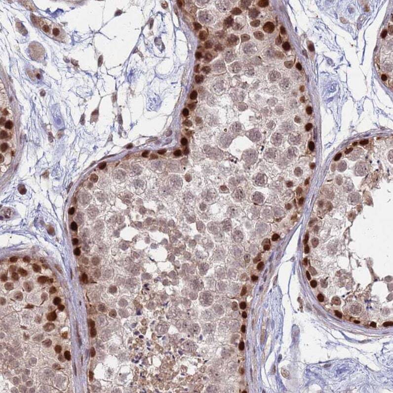
- Immunohistochemical analysis of TRMT6 in human testis using TRMT6 Polyclonal Antibody (Product # PA5-61409) shows strong nuclear positivity in cells in seminiferous ducts.
Supportive validation
- Submitted by
- Invitrogen Antibodies (provider)
- Main image

- Experimental details
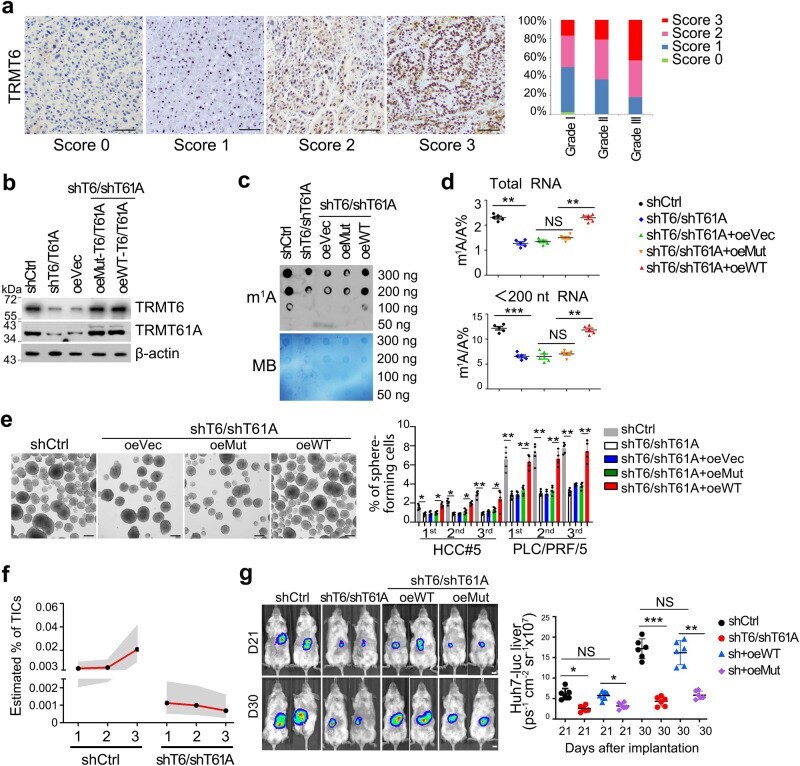
- Fig. 2 TRMT6/TRMT61A-mediated m 1 A modification drives self-renewal of liver CSCs and tumourigenesis. a Representative immunohistochemistry images of TRMT6 protein levels (brown) in the cohort of 191 HCC samples. n = 3 independent experiments. Score 0 (0-5% positive cells/tissue section) represents TRMT6 staining that is considered negative; score 1 (5-15% positive cells/section) represents TRMT6 staining that is considered week positive, whereas scores 2 (15-35% positive cells/section) and 3 (>35% positive cells/section) represent TRMT6 staining that is considered positive. Grade I ( n = 56), Grade II ( n = 87), Grade III ( n = 48). Scale bar, 100 mum. b Western blotting confirmation of TRMT6/TRMT61A ( T6/T61A ) depletion, and co-infection of WT or TRMT6 R377L / TRMT61A D181A (Mut) constructs in TRMT6/TRMT61A depleted cells. These experiments were repeated three times. oe overexpression, Vec vector. n = 3 biologically independent samples. c Global m 1 A levels were detected in shCtrl, TRMT6/TRMT61A depletion, and co-infection of WT or Mut- TRMT6/TRMT61A in TRMT6/TRMT61A depleted liver CSCs using dot blot assay. Data were repeated five times. n = 5 biologically independent samples. d LC-MS/MS quantification of m 1 A levels in total RNA and tRNA (