Explore
 Validate
 Learn
Learn Western blot
 Immunocytochemistry
Antibody data
- Antibody Data
- Antigen structure
- References [1]
- Comments [0]
- Validations
- Immunocytochemistry [3]
- Immunohistochemistry [1]
- Other assay [1]
Submit
Validation data
Reference
Comment
Report error
- Product number
- PA5-21672 - Provider product page

- Provider
- Invitrogen Antibodies
- Product name
- AKR1C1 Polyclonal Antibody
- Antibody type
- Polyclonal
- Antigen
- Recombinant full-length protein
- Description
- Recommended positive controls: A431, HeLa, HepG2, A549, mouse liver. Predicted reactivity: Pig (83%), Rhesus Monkey (95%). Store product as a concentrated solution. Centrifuge briefly prior to opening the vial.
- Reactivity
- Human, Mouse
- Host
- Rabbit
- Isotype
- IgG
- Vial size
- 100 μL
- Concentration
- 0.6 mg/mL
- Storage
- Store at 4°C short term. For long term storage, store at -20°C, avoiding freeze/thaw cycles.
Submitted references Regulation of uterine function during estrous cycle, anestrus phase and pregnancy by steroids in red deer (Cervus elaphus L.).
Kotlarczyk AM, Grzyb M, Korzekwa AJ
Scientific reports 2021 Oct 11;11(1):20109
Scientific reports 2021 Oct 11;11(1):20109
No comments: Submit comment
Supportive validation
- Submitted by
- Invitrogen Antibodies (provider)
- Main image

- Experimental details
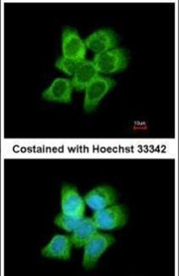
- Immunofluorescent analysis of AKR1C1 in methanol-fixed A431 cells using an AKR1C1 polyclonal antibody (Product # PA5-21672) at a 1:200 dilution.
- Submitted by
- Invitrogen Antibodies (provider)
- Main image

- Experimental details
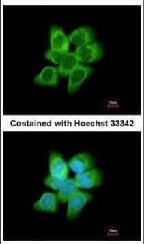
- Immunofluorescence analysis of methanol-fixed A431, using AKR1C1 antibody (Product # PA5-21672) at 1:200 dilution.
- Submitted by
- Invitrogen Antibodies (provider)
- Main image

- Experimental details
- Immunofluorescence analysis of methanol-fixed A431, using AKR1C1 antibody (Product # PA5-21672) at 1:200 dilution.

Supportive validation
- Submitted by
- Invitrogen Antibodies (provider)
- Main image

- Experimental details
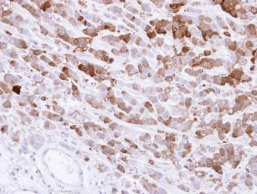
- Immunohistochemical analysis of paraffin-embedded MDA-MB-468 xenograft , using AKR1C1 (Product # PA5-21672) antibody at 1:500 dilution. Antigen Retrieval: Citrate buffer, pH 6.0, 15 min.
Supportive validation
- Submitted by
- Invitrogen Antibodies (provider)
- Main image
- Experimental details
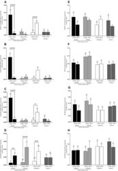
- Figure 1 mRNA and protein expression of AKR1C1 ( A , E ), P450 ( B , F ), 3beta-HSD ( C , G ) and 17beta-HSD ( D , H ) in uterine tissues (endometrium and myometrium) on 4th and 13th day of estrous cycle, in pregnancy and anestrus phase. Data were normalized against GAPDH for mRNA expression and against beta-actin (ACTB) for proteins expression. Each bar represents one experimental group with SEM. Statistical differences were analyzed by two-way analysis (ANOVA) of variance followed by the Bonferroni post hoc test using GraphPad PRISM (Version 8.3.0). The lowest statistical significance was P < 0.05. Asterisks indicate statistical differences between endometrium and myometrium (* P < 0.1; ** P < 0.01; *** P < 0.001; **** P < 0.0001). Different letters indicate statistical differences ( P < 0.05) between the experimental groups throughout endometrium (A, B) and myometrium (a-b) respectively.