 Explore
 Validate
 Learn
Learn Western blot
 Immunohistochemistry
Antibody data
- Antibody Data
- Antigen structure
- References [1]
- Comments [0]
- Validations
- Immunohistochemistry [1]
- Other assay [2]
Submit
Validation data
Reference
Comment
Report error
- Product number
- PA5-41677 - Provider product page

- Provider
- Invitrogen Antibodies
- Product name
- GLIS3 Polyclonal Antibody
- Antibody type
- Polyclonal
- Antigen
- Synthetic peptide
- Description
- Peptide sequence: PPASQVSTAC NQISPSLQRA MNAANLNIPP SDTRSLISRE SLASTTLSLT Sequence homology: Dog: 100%; Guinea Pig: 100%; Horse: 100%; Human: 100%; Mouse: 93%; Rabbit: 100%; Rat: 100%
- Reactivity
- Human
- Host
- Rabbit
- Isotype
- IgG
- Vial size
- 100 μL
- Concentration
- 1 mg/mL
- Storage
- -20°C, Avoid Freeze/Thaw Cycles
Submitted references Cryptotanshinone Protects Cartilage against Developing Osteoarthritis through the miR-106a-5p/GLIS3 Axis.
Ji Q, Qi D, Xu X, Xu Y, Goodman SB, Kang L, Song Q, Fan Z, Maloney WJ, Wang Y
Molecular therapy. Nucleic acids 2018 Jun 1;11:170-179
Molecular therapy. Nucleic acids 2018 Jun 1;11:170-179
No comments: Submit comment
Supportive validation
- Submitted by
- Invitrogen Antibodies (provider)
- Main image
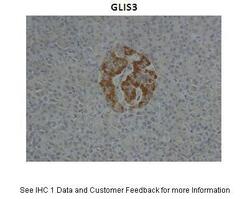
- Experimental details
- Immunohistochemistry analysis of human pancreas cells using an anti-GLIS3 polyclonal antibody (Product # PA5-41677). Primary Antibody Dilution:1:10; Secondary Antibody: Anti-rabbit HRP; Secondary Antibody Dilution: 1:1000.
Supportive validation
- Submitted by
- Invitrogen Antibodies (provider)
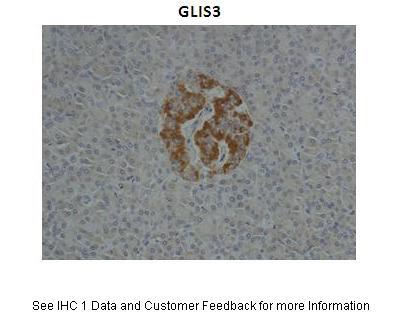
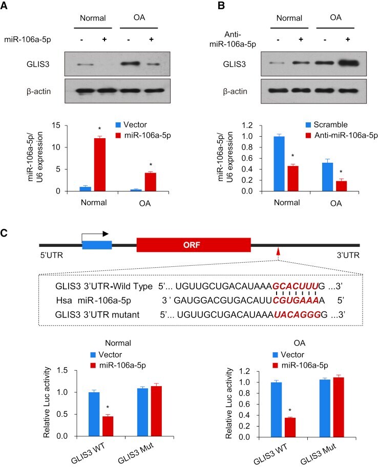
- Main image

- Experimental details
- Figure 3 miR-106a-5p Inhibits GLIS3 Expression by Targeting its 3'-UTR (A and B) Immunoblot analysis of chondrocytes transfected with miR-106a-5p (A) or anti-miR-106a-5p (B). The histograms shown under the immunoblot graphs reveal corresponding miR-106a-5p expression levels. (C) miRNA luciferase reporter assay in human chondrocytes cotransfected with wild-type or mutated GLIS3 reporter and miR-106a-5p. The top panel shows the wild-type and mutant forms of the putative miR-106a-5p target sequences in the GLIS3 3'-UTR. Bold and red italicized font indicate the putative miR-106a-5p binding sites within the human GLIS3 3'-UTR. GLIS3 WT, wild-type GLIS3 3'-UTR; GLIS3 Mut, mutated GLIS3 3'UTR. Each bar represents the mean +- SD of at least three independent experiments performed in triplicate. *p < 0.05, **p < 0.01.
- Submitted by
- Invitrogen Antibodies (provider)
- Main image
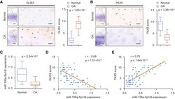
- Experimental details
- Figure 5 Expression of GLIS3, PAX5, and miR-106a-5p in OA Patients (A) Representative immunohistochemistry assay of GLIS3 in normal and OA cartilage tissues. Scale bar, left, 500 mum; right, 50 mum. The scores of GLIS3 in normal and OA cartilage tissues based on the immunohistochemistry assay are shown. (B) Representative immunohistochemistry assay of PAX5 in normal and OA cartilage tissues. Scale bar, left, 500 mum; right, 50 mum. The scores of PAX5 in normal and OA cartilage tissues based on the immunohistochemistry assay. (C) qRT-PCR measurement of miR-106a-5p in normal and OA cartilage tissues. Data are expressed as the mean +- SD. (D and E) The relationship among GLIS3 (D), PAX5 (E), and miR-106a-5p expression was determined through Pearson's chi2 analysis.
