Explore
 Validate
 Learn
Learn Western blot
 Immunocytochemistry
Antibody data
- Antibody Data
- Antigen structure
- References [3]
- Comments [0]
- Validations
- Immunocytochemistry [2]
- Immunoprecipitation [1]
- Immunohistochemistry [4]
- Other assay [4]
Submit
Validation data
Reference
Comment
Report error
- Product number
- PA5-53023 - Provider product page

- Provider
- Invitrogen Antibodies
- Product name
- VAPB Polyclonal Antibody
- Antibody type
- Polyclonal
- Antigen
- Recombinant protein fragment
- Description
- Immunogen sequence: VWKEAKPEDL MDSKLRCVFE LPAENDKPHD VEINKIISTT ASKTETPIVS KSLSSSLDDT EVKKVMEECK RLQGEVQRLR EENKQFKEED GLRMRKTVQS NSPISALAPT GK Highest antigen sequence identity to the following orthologs: Mouse - 82%, Rat - 79%.
- Reactivity
- Human, Mouse, Rat
- Host
- Rabbit
- Isotype
- IgG
- Vial size
- 100 μL
- Concentration
- 0.3 mg/mL
- Storage
- Store at 4°C short term. For long term storage, store at -20°C, avoiding freeze/thaw cycles.
Submitted references Role of Vesicle-Associated Membrane Protein-Associated Proteins (VAP) A and VAPB in Nuclear Egress of the Alphaherpesvirus Pseudorabies Virus.
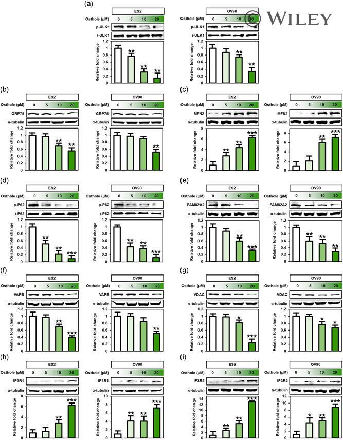
Osthole interacts with an ER-mitochondria axis and facilitates tumor suppression in ovarian cancer.
Stigmasterol Causes Ovarian Cancer Cell Apoptosis by Inducing Endoplasmic Reticulum and Mitochondrial Dysfunction.
Dorsch AD, Hölper JE, Franzke K, Zaeck LM, Mettenleiter TC, Klupp BG
Viruses 2021 Jun 10;13(6)
Viruses 2021 Jun 10;13(6)
Osthole interacts with an ER-mitochondria axis and facilitates tumor suppression in ovarian cancer.
Bae H, Lee JY, Song J, Song G, Lim W
Journal of cellular physiology 2021 Feb;236(2):1025-1042
Journal of cellular physiology 2021 Feb;236(2):1025-1042
Stigmasterol Causes Ovarian Cancer Cell Apoptosis by Inducing Endoplasmic Reticulum and Mitochondrial Dysfunction.
Bae H, Song G, Lim W
Pharmaceutics 2020 May 28;12(6)
Pharmaceutics 2020 May 28;12(6)
No comments: Submit comment
Supportive validation
- Submitted by
- Invitrogen Antibodies (provider)
- Main image

- Experimental details
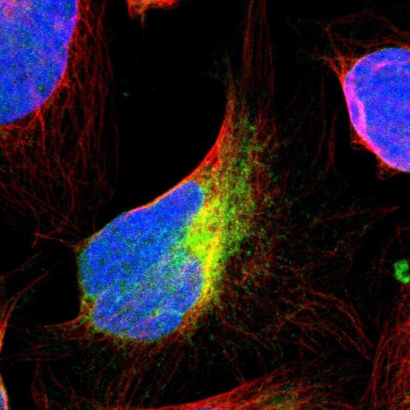
- Immunofluorescent staining of VAPB in human cell line U-2 OS shows positivity in endoplasmic reticulum. Samples were probed using a VAPB Polyclonal Antibody (Product # PA5-53023).

- Submitted by
- Invitrogen Antibodies (provider)
- Main image

- Experimental details
- Immunofluorecent analysis of VAPB in human cell line U-2 OS using VAPB Polyclonal Antibody (Product # PA5-53023). Staining shows positivity in endoplasmic reticulum.
Supportive validation
- Submitted by
- Invitrogen Antibodies (provider)
- Main image

- Experimental details
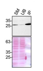
- Immunoprecipitation of VAPB was performed on HeLa cell lysates. Antibody-bead conjugates were prepared by adding 1 µg of VAPB polyclonal antibody (Product # PA5-53023) with 30 µL of protein A-Sepharose beads and rocked overnight at 4°C. 1 mg of lysate was incubated with an antibody-bead conjugate for 2 hours at 4°C. Following centrifugation and multiple washes, 10% starting material (SM), 10% unbound fraction (UB) and immunoprecipitated fraction (IP) were processed for immunoblot using a different VAPB antibody. Ponceau stained transfer of blot is shown. Data courtesy of YCharOS Inc., an open science company with the mission of characterizing commercially available antibodies using knockout validation.

Supportive validation
- Submitted by
- Invitrogen Antibodies (provider)
- Main image

- Experimental details
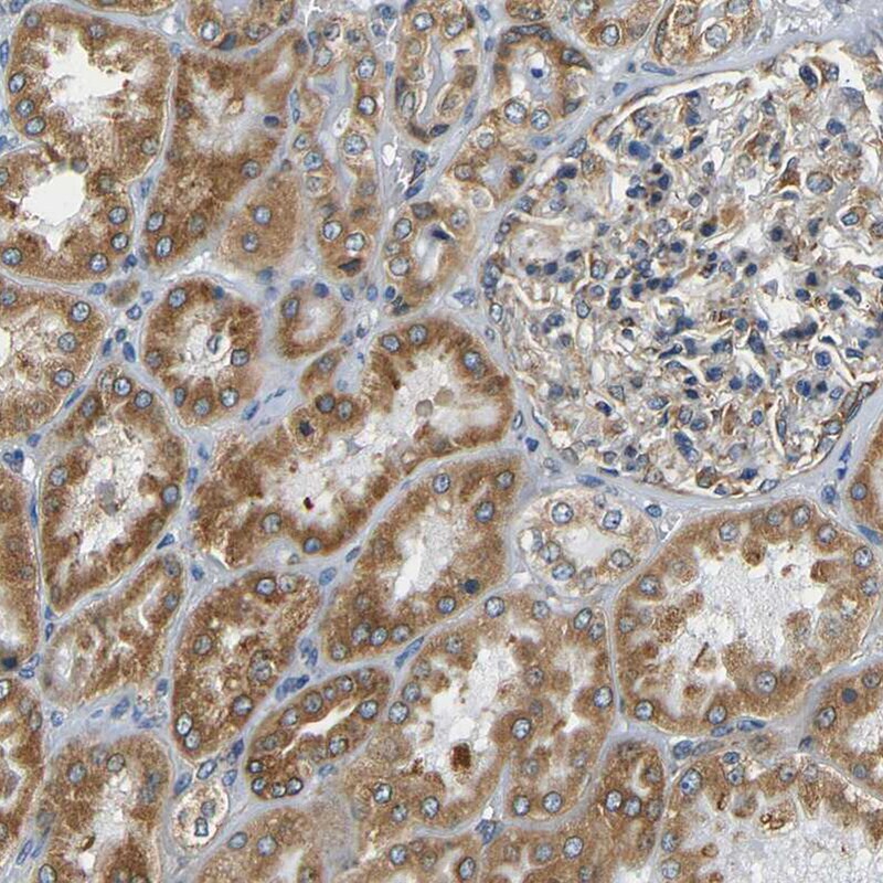
- Immunohistochemical staining of VAPB in human kidney using a VAPB Polyclonal Antibody (Product # PA5-53023) shows moderate cytoplasmic positivity in cells in tubules.
- Submitted by
- Invitrogen Antibodies (provider)
- Main image

- Experimental details
- Immunohistochemical staining of VAPB in human liver using a VAPB Polyclonal Antibody (Product # PA5-53023) shows moderate cytoplasmic positivity in hepatocytes.

- Submitted by
- Invitrogen Antibodies (provider)
- Main image
- Experimental details
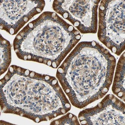
- Immunohistochemical staining of VAPB in human small intestine using a VAPB Polyclonal Antibody (Product # PA5-53023) shows strong cytoplasmic positivity in glandular cells.

- Submitted by
- Invitrogen Antibodies (provider)
- Main image
- Experimental details
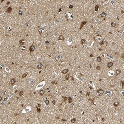
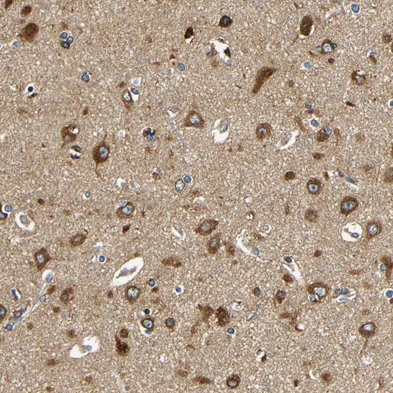
- Immunohistochemical staining of VAPB in human cerebral cortex using a VAPB Polyclonal Antibody (Product # PA5-53023) shows strong cytoplasmic positivity in neuronal cells.
Supportive validation
- Submitted by
- Invitrogen Antibodies (provider)
- Main image
- Experimental details
- Immunoprecipitation of VAPB was performed on HeLa cell lysates. Antibody-bead conjugates were prepared by adding 1 µg of VAPB polyclonal antibody (Product # PA5-53023) with 30 µL of protein A-Sepharose beads and rocked overnight at 4°C. 1 mg of lysate was incubated with an antibody-bead conjugate for 2 hours at 4°C. Following centrifugation and multiple washes, 10% starting material (SM), 10% unbound fraction (UB) and immunoprecipitated fraction (IP) were processed for immunoblot using a different VAPB antibody. Ponceau stained transfer of blot is shown. Data courtesy of YCharOS Inc., an open science company with the mission of characterizing commercially available antibodies using knockout validation.

- Submitted by
- Invitrogen Antibodies (provider)
- Main image

- Experimental details
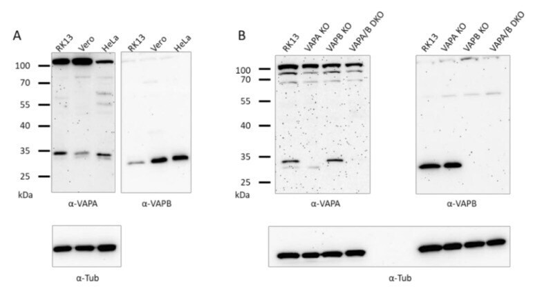
- Figure 2 Immunoblots showing VAPA and VAPB in RK13, Vero and HeLa cells and the absence of VAPA and/or VAPB in the corresponding RK13 KO and DKO cell lines. Proteins in lysates of RK13, Vero and HeLa cells ( A ) or RK13 and the single as well as the double VAP KO cell lines ( B ) were separated on SDS-10 % polyacrylamide gels, and parallel blots were incubated with polyclonal sera against VAPA or VAPB. Masses of marker proteins are given on the left in kDa. As loading control, parallel ( A ) or the same blots were (re-)probed with anti-alpha-tubulin.
- Submitted by
- Invitrogen Antibodies (provider)
- Main image
- Experimental details
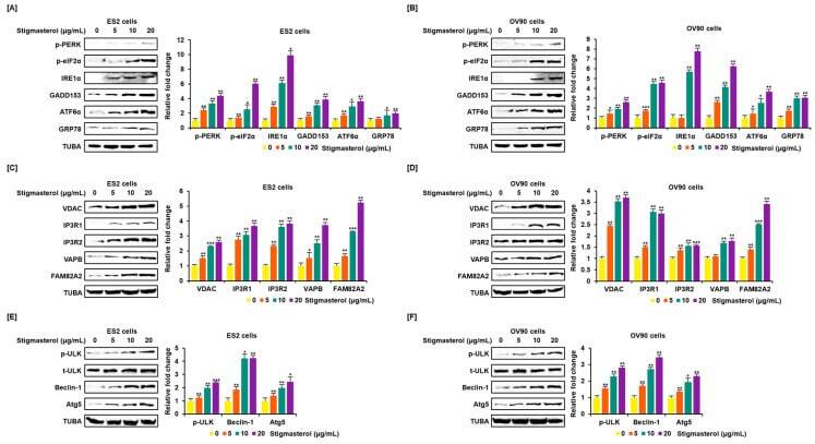
- Figure 4 Increased expression of endoplasmic reticulum (ER) stress sensor proteins and ER-mitochondrial axis proteins due to stigmasterol treatment in ovarian cancer cells. ( A , B ) Immunoblots representing the expression of unfolded protein response (UPR) proteins following stigmasterol treatments (0, 5, 10, and 20 mug/mL). TUBA was used as a control and is shown at the bottom of the figure. ( C , D ) Immunoblots representing the expression of ER-mitochondrial axis proteins following stigmasterol treatments (0, 5, 10, and 20 mug/mL). TUBA was used as a control and is shown at the bottom of the figure. ( E , F ) Immunoblots representing the expression of autophagy proteins following stigmasterol treatment. TUBA was used as a control and is shown at the bottom of the figure.
- Submitted by
- Invitrogen Antibodies (provider)
- Main image
- Experimental details
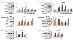
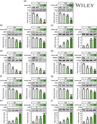
- 8 Figure Osthole influenced intracellular mechanisms related to endoplasmic reticulum (ER)-mitochondrial functions. Expression of ER-mitochondrial function targets such as (a) ULK1, (b) GRP75, (c) MFN2, (d) P62, (e) FAM82A2, (f) VAPB, (g) VDAC, (h) IP3R1, and (i) IP3R2 was analyzed by western blot analysis in ES2 and OV90 cells treated with various doses of osthole (0, 5, 10, and 20 muM). Intensity of the target protein was detected and analyzed relative to total protein or alpha-tubulin protein. *** p < .001, ** p < .01, and * p < .05 indicate significant differences compared to vehicle-treated cells. IP3R1, inositol 1,4,5-trisphosphate (IP3) receptor type 1; IP3, inositol trisphosphate; MFN2, mitofusin-2; VAPB, vesicle-associated membrane protein-associated protein B/C; VDAC, voltage-dependent anion channel