Explore
 Validate
 Learn
LearnPA5-36572
antibody from Invitrogen Antibodies
Targeting: AKR1C2
BABP, DD, DD2, DDH2, HAKRD, MCDR2, TDD
 Western blot
 Immunohistochemistry
 Other assay
Antibody data
- Antibody Data
- Antigen structure
- References [2]
- Comments [0]
- Validations
- Other assay [1]
Submit
Validation data
Reference
Comment
Report error
- Product number
- PA5-36572 - Provider product page

- Provider
- Invitrogen Antibodies
- Product name
- AKR1C2 Polyclonal Antibody
- Antibody type
- Polyclonal
- Antigen
- Synthetic peptide
- Description
- This antibody detects endogenous protein at a molecular weight of 37 kDa. Purity is >95% by SDS-PAGE.
- Reactivity
- Human, Mouse, Rat
- Host
- Rabbit
- Isotype
- IgG
- Vial size
- 100 μL
- Concentration
- 1 mg/mL
- Storage
- Store at 4°C short term. For long term storage, store at -20°C, avoiding freeze/thaw cycles.
Submitted references Endothelin-1 induces changes in the expression levels of steroidogenic enzymes and increases androgen receptor and testosterone production in the PC3 prostate cancer cell line.
Silencing of the transcriptional factor ZEB1 alters the steroidogenic pathway, and increases the concentration of testosterone and DHT in DU145 cells.
Torres MJ, López-Moncada F, Herrera D, Indo S, Lefian A, Llanos P, Tapia J, Castellón EA, Contreras HR
Oncology reports 2021 Aug;46(2)
Oncology reports 2021 Aug;46(2)
Silencing of the transcriptional factor ZEB1 alters the steroidogenic pathway, and increases the concentration of testosterone and DHT in DU145 cells.
Herrera D, Orellana-Serradell O, Villar P, Torres MJ, Paciucci R, Castellón EA, Contreras HR
Oncology reports 2019 Feb;41(2):1275-1283
Oncology reports 2019 Feb;41(2):1275-1283
No comments: Submit comment
Supportive validation
- Submitted by
- Invitrogen Antibodies (provider)
- Main image

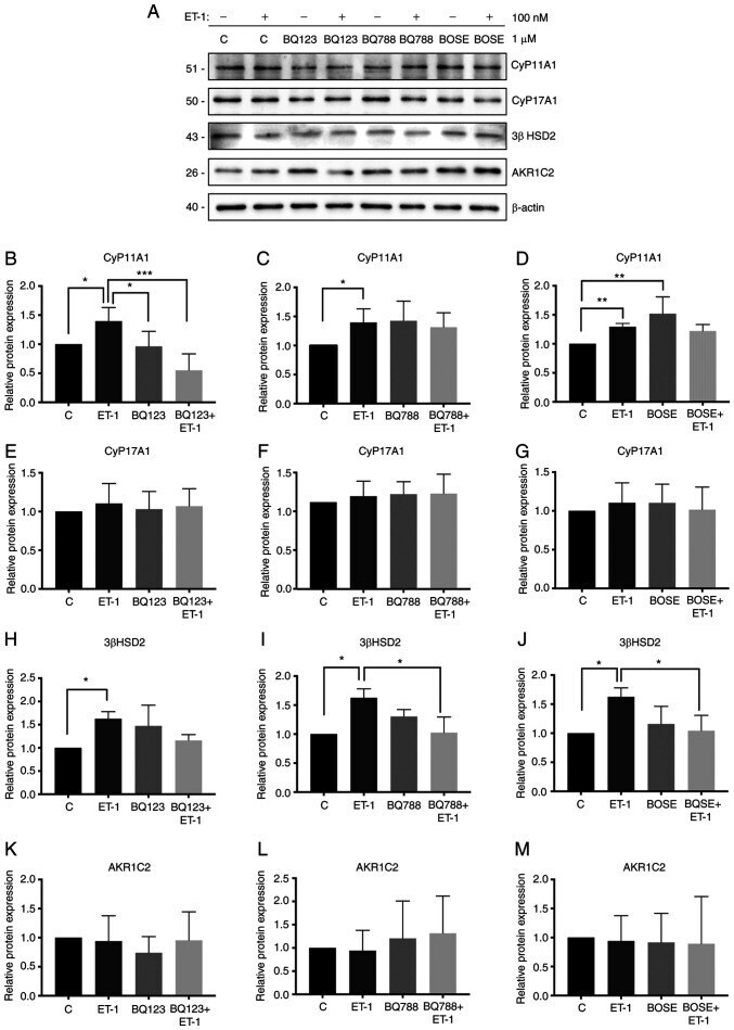
- Experimental details
- Figure 4. Effect of ET-1 on the expression levels of steroidogenic enzymes. (A) Lysed PC3 cells were analyzed by western blotting and membranes were incubated with antibodies against CyP11A1, CyP17A1, 3beta HSD2, AKR1C2 and beta-actin. (B) Protein expression levels of CyP11A1 in the presence or absence of BQ123. (C) Protein expression levels of CyP11A1 in the presence or absence of BQ788. (D) Protein expression levels of CyP11A1 in the presence or absence of BOSE. (E) Protein expression levels of CyP17A1 in the presence or absence of BQ123. (F) Protein expression levels of CyP17A1 in the presence or absence of BQ788. (G) Protein expression levels of CyP17A1 in the presence or absence of BOSE. (H) Protein expression levels of 3beta HSD2 in the presence or absence of BQ123. (I) Protein expression levels of 3beta HSD2 in the presence or absence of BQ788. (J) Protein expression levels of 3beta HSD2 in the presence or absence of BOSE. (K) Protein expression levels of AKR1C2 in the presence or absence of BQ123. (L) Protein expression levels of AKR1C2 in the presence or absence of BQ788. (M) Protein expression levels of AKR1C2 in the presence or absence of BOSE. Quantification was normalized to beta-actin and control PC3 cells. Data are presented as the mean +- SD (n=3 independent experiments). *P