 Explore
 Validate
 Learn
Learn Western blot
 Immunocytochemistry
Antibody data
- Antibody Data
- Antigen structure
- References [3]
- Comments [0]
- Validations
- Western blot [2]
- Immunohistochemistry [9]
Submit
Validation data
Reference
Comment
Report error
- Product number
- NBP1-90264 - Provider product page

- Provider
- Novus Biologicals
- Proper citation
- Novus Cat#NBP1-90264, RRID:AB_11007848
- Product name
- Rabbit Polyclonal Aconitase 2 Antibody
- Antibody type
- Polyclonal
- Description
- Immunogen affinity purified. Specificity of human, mouse, rat Aconitase 2 antibody verified on a Protein Array containing target protein plus 383 other non-specific proteins.
- Reactivity
- Human, Mouse, Rat
- Host
- Rabbit
- Isotype
- IgG
- Vial size
- 0.1 ml
- Storage
- Store at 4C short term. Aliquot and store at -20C long term. Avoid freeze-thaw cycles.
Submitted references Exosomal miRNA Let-7 from Menstrual Blood-Derived Endometrial Stem Cells Alleviates Pulmonary Fibrosis through Regulating Mitochondrial DNA Damage.
STED super-resolution microscopy of clinical paraffin-embedded human rectal cancer tissue.
Mitochondrial aconitase knockdown attenuates paraquat-induced dopaminergic cell death via decreased cellular metabolism and release of iron and H₂O₂.
Sun L, Zhu M, Feng W, Lin Y, Yin J, Jin J, Wang Y
Oxidative medicine and cellular longevity 2019;2019:4506303
Oxidative medicine and cellular longevity 2019;2019:4506303
STED super-resolution microscopy of clinical paraffin-embedded human rectal cancer tissue.
Ilgen P, Stoldt S, Conradi LC, Wurm CA, Rüschoff J, Ghadimi BM, Liersch T, Jakobs S
PloS one 2014;9(7):e101563
PloS one 2014;9(7):e101563
Mitochondrial aconitase knockdown attenuates paraquat-induced dopaminergic cell death via decreased cellular metabolism and release of iron and H₂O₂.
Cantu D, Fulton RE, Drechsel DA, Patel M
Journal of neurochemistry 2011 Jul;118(1):79-92
Journal of neurochemistry 2011 Jul;118(1):79-92
No comments: Submit comment
Supportive validation
- Submitted by
- Novus Biologicals (provider)
- Main image
- Experimental details
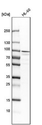
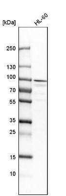
- Western Blot: Aconitase 2 Antibody [NBP1-90264] - Analysis in human cell line HL-60.
- Submitted by
- Novus Biologicals (provider)
- Main image

- Experimental details
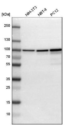
- Western Blot: Aconitase 2 Antibody [NBP1-90264] - Western blot analysis in mouse cell line NIH-3T3, rat cell line NBT-II and rat cell line pC12.
Supportive validation
- Submitted by
- Novus Biologicals (provider)
- Main image
- Experimental details
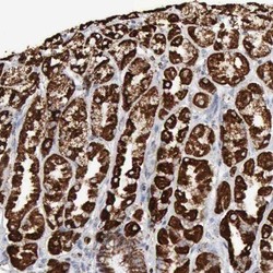
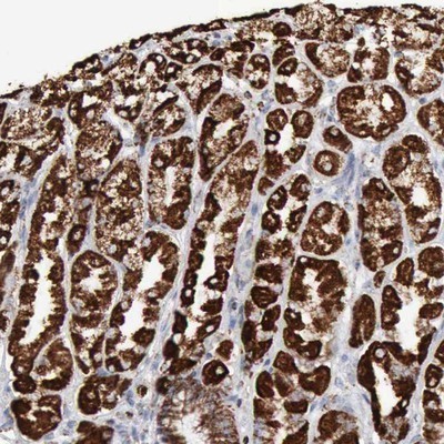
- Immunohistochemistry-Paraffin: Aconitase 2 Antibody [NBP1-90264] - Staining of human stomach shows strong cytoplasmic positivity in glandular cells.
- Submitted by
- Novus Biologicals (provider)
- Main image
- Experimental details
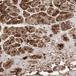
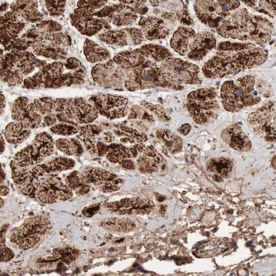
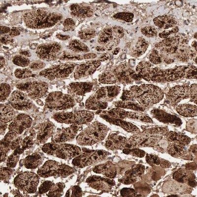
- Immunohistochemistry-Paraffin: Aconitase 2 Antibody [NBP1-90264] - Staining of human heart muscle shows high expression.
- Submitted by
- Novus Biologicals (provider)
- Main image
- Experimental details
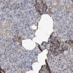
- Immunohistochemistry-Paraffin: Aconitase 2 Antibody [NBP1-90264] - Staining of human pancreas shows low expression as expected.

- Submitted by
- Novus Biologicals (provider)
- Main image
- Experimental details
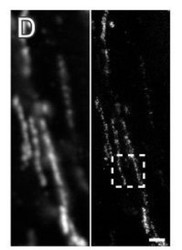
- Immunohistochemistry: Aconitase 2 Antibody [NBP1-90264] - STED super-resolution microscopy of mitochondria in the rectal Muscularis externa demonstrates high structural preservation of the stored paraffin-embedded tissue. STED recordings were performed on 2 um thick dewaxed sections cut along the longitudinal axis of the rectum. STED images of tissue sections decorated with antisera against aconitase. In each panel the confocal (left) and the corresponding STED image (right) is displayed. Magnification of the STED image as indicated by a dashed square. Scale bar: 200 nm. Image collected and cropped by CiteAb from the following publication (http://dx.plos.org/10.1371/journal.pone.0101563), licensed under a CC-BY licence.

- Submitted by
- Novus Biologicals (provider)
- Main image
- Experimental details
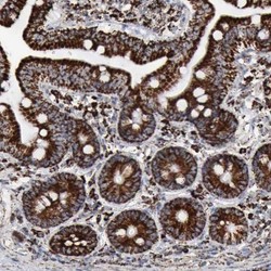
- Immunohistochemistry-Paraffin: Aconitase 2 Antibody [NBP1-90264] - Staining of human duodenum shows strong strong granular cytoplasmic positivity in glandular cells.

- Submitted by
- Novus Biologicals (provider)
- Main image
- Experimental details
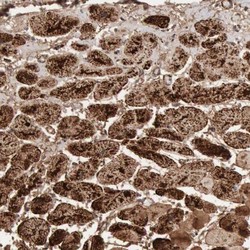
- Immunohistochemistry-Paraffin: Aconitase 2 Antibody [NBP1-90264] - Staining of human heart muscle shows strong granular cytoplasmic positivity in cardiomyocytes.
- Submitted by
- Novus Biologicals (provider)
- Main image
- Experimental details
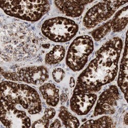
- Immunohistochemistry-Paraffin: Aconitase 2 Antibody [NBP1-90264] - Staining of human kidney shows strong strong granular cytoplasmic positivity in cells in tubules.

- Submitted by
- Novus Biologicals (provider)
- Main image
- Experimental details
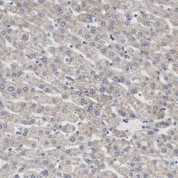
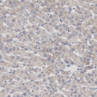
- Immunohistochemistry-Paraffin: Aconitase 2 Antibody [NBP1-90264] - Staining of human liver shows very weak positivity in hepatocytes as expected.
- Submitted by
- Novus Biologicals (provider)
- Main image
- Experimental details
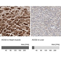
- Immunohistochemistry-Paraffin: Aconitase 2 Antibody [NBP1-90264] - Staining in human heart muscle and liver tissues using NBP1-90264 antibody. Corresponding ACO2 RNA-seq data are presented for the same tissues.
