Explore
 Validate
 Learn
LearnHPA030196
antibody from Atlas Antibodies
Targeting: PDXK
C21orf124, C21orf97, FLJ21324, FLJ31940, MGC15873, PKH, PNK, PRED79
 Western blot
 Immunocytochemistry
Antibody data
- Antibody Data
- Antigen structure
- References [1]
- Comments [0]
- Validations
- Immunocytochemistry [1]
- Immunohistochemistry [1]
Submit
Validation data
Reference
Comment
Report error
- Product number
- HPA030196 - Provider product page

- Provider
- Atlas Antibodies
- Proper citation
- Atlas Antibodies Cat#HPA030196, RRID:AB_10599735
- Product name
- Anti-PDXK
- Antibody type
- Polyclonal
- Description
- Polyclonal Antibody against Human PDXK, Gene description: pyridoxal (pyridoxine, vitamin B6) kinase, Alternative Gene Names: C21orf124, C21orf97, FLJ21324, FLJ31940, MGC15873, PKH, PNK, PRED79, Validated applications: ICC, IHC, WB, Uniprot ID: O00764, Storage: Store at +4°C for short term storage. Long time storage is recommended at -20°C.
- Reactivity
- Human
- Host
- Rabbit
- Conjugate
- Unconjugated
- Isotype
- IgG
- Vial size
- 100 µl
- Concentration
- 0.3 mg/ml
- Storage
- Store at +4°C for short term storage. Long time storage is recommended at -20°C.
- Handling
- The antibody solution should be gently mixed before use.
Submitted references New Insights Into Pyridoxal Kinase Inhibitors and Their Antileukemic Effects
Banerjee P, Singh T, Qamar I
Cureus 2023
Cureus 2023
No comments: Submit comment
Supportive validation
- Submitted by
- Atlas Antibodies (provider)
- Main image
- Experimental details
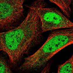
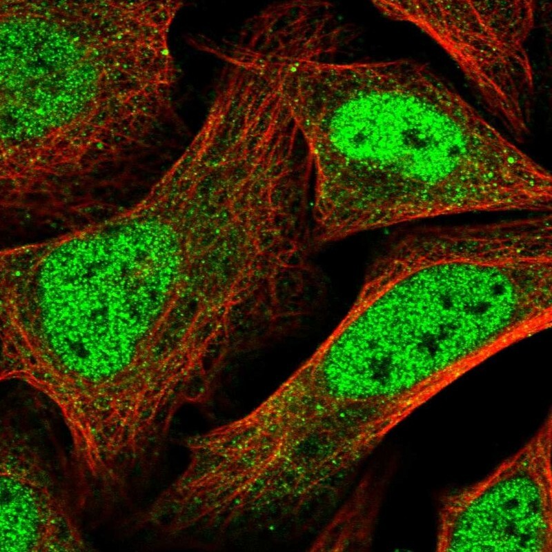
- Immunofluorescent staining of human cell line U-2 OS shows localization to nucleoplasm.
- Sample type
- Human
Supportive validation
- Submitted by
- Atlas Antibodies (provider)
- Enhanced method
- Orthogonal validation
- Main image

- Experimental details
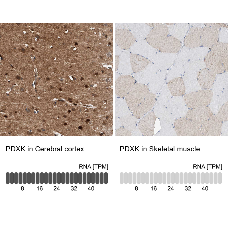
- Immunohistochemistry analysis in human cerebral cortex and skeletal muscle tissues using HPA030196 antibody. Corresponding PDXK RNA-seq data are presented for the same tissues.
- Sample type
- Human
- Protocol
- Protocol