Explore
 Validate
 Learn
Learn Western blot
Antibody data
- Antibody Data
- Antigen structure
- References [0]
- Comments [0]
- Validations
- Western blot [1]
- Immunocytochemistry [1]
- Flow cytometry [1]
Submit
Validation data
Reference
Comment
Report error
- Product number
- MA1-179 - Provider product page

- Provider
- Invitrogen Antibodies
- Product name
- eIF5A Monoclonal Antibody (4E10G8)
- Antibody type
- Monoclonal
- Antigen
- Purifed from natural sources
- Description
- MA1-179 detects EIF5A from human samples.
- Antibody clone number
- 4E10G8
- Concentration
- 1 mg/mL
No comments: Submit comment
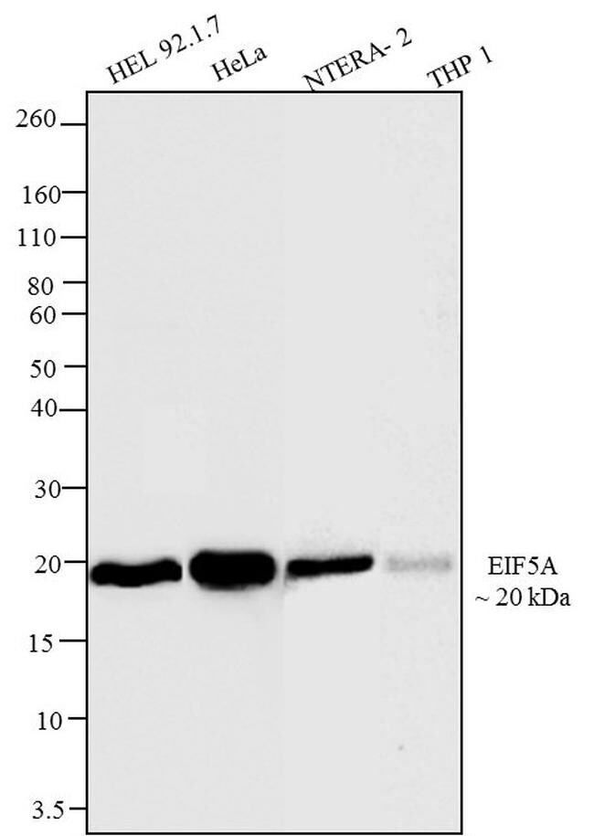
Supportive validation
- Submitted by
- Invitrogen Antibodies (provider)
- Main image

- Experimental details
- Western blot analysis of EIF5A was performed on whole cell extracts (30 µg lysate) of HEL 92.1.7 (Lane 1), HeLa (Lane 2), NTERA-2 (Lane 3) and THP1 (Lane 4). Protein samples were electrophoresed using a Novex® NuPAGE® 10 % Bis-Tris gel (Product # NP0302BOX), XCell SureLock™ Electrophoresis System (Product # EI0002), and Novex® Sharp Pre-Stained Protein Standard (Product # LC5800). Resolved proteins were then transferred onto a nitrocellulose membrane with the iBlot® 2 Dry Blotting System (Product # IB21001), and blocked with 5% Milk in PBST. The blots were probed with a EIF5A Mouse Monoclonal Antibody (Product # MA1-179, 1-2 µg/mL) followed by a Goat anti-Mouse IgG (H+L) Superclonal™ Secondary Antibody, HRP conjugate (Product # A28177, 0.4 µg/mL, 1:2500 dilution). Chemiluminescent detection was performed using Pierce™ ECL Western Blotting Substrate (Product # 32106). EIF5A was detected at approximately 20kDa across cell lines tested.
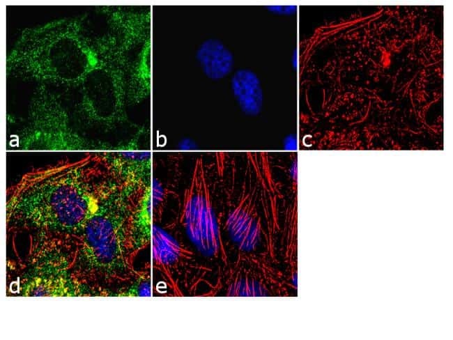
Supportive validation
- Submitted by
- Invitrogen Antibodies (provider)
- Main image
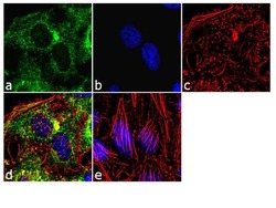
- Experimental details
- Immunofluorescent analysis of EIF5A was performed using 70% confluent log phase HeLa cells. The cells were fixed with 4% paraformaldehyde for 10 minutes, permeabilized with 0.1% Triton™ X-100 for 10 minutes, and blocked with 1% BSA for 1 hour at room temperature. The cells were then labeled with a EIF5A Mouse Monoclonal Antibody (Product # MA1-179) at 2 µg/mL in 0.1% BSA for 3 hours at room temperature, and then labeled with a Goat anti-Mouse IgG (H+L) Superclonal™ Secondary Antibody, Alexa Fluor® 488 conjugate (Product # A28175) at a dilution of 1:2000 for 45 minutes at room temperature (Panel a: green). Nuclei (Panel b: blue) were stained with SlowFade® Gold Antifade Mountant with DAPI (Product # S36938). F-actin (Panel c: red) was stained with Alexa Fluor® 555 Rhodamine Phalloidin (Product # R415, 1:300). Panel d represents the merged image showing cytoplasmic localization. Panel e shows the no primary antibody control. The images were captured at 60X magnification.
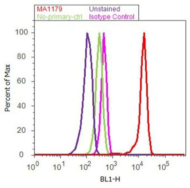
Supportive validation
- Submitted by
- Invitrogen Antibodies (provider)
- Main image

- Experimental details
- Flow cytometry analysis of EIF5A was done on HeLa cells. Cells were fixed with 70% ethanol for 10 minutes, permeabilized with 0.25% Triton™ X-100 for 20 minutes, and blocked with 5% BSA for 30 minutes at room temperature. Cells were then labeled with a EIF5A Mouse Monoclonal Antibody (Product # MA1-179, red histogram) or a mouse isotype control (pink histogram) at 3-5 µg/million cells in 2.5% BSA. After incubation at room temperature for 2 hours, the cells were then labeled with a Rabbit anti-Mouse IgG (H+L) Secondary Antibody, Alexa Fluor® 488 conjugate (Product # A-11059) at a dilution of 1:400 for 30 minutes at room temperature. The representative 10,000 cells were acquired and analyzed for each sample using an Attune® Acoustic Focusing Cytometer. The purple histogram represents unstained control cells and the green histogram represents no primary antibody control.