Explore
 Validate
 Learn
Learn Western blot
 Immunocytochemistry
Antibody data
- Antibody Data
- Antigen structure
- References [1]
- Comments [0]
- Validations
- Western blot [1]
Submit
Validation data
Reference
Comment
Report error
- Product number
- PB9481 - Provider product page

- Provider
- Boster Biological Technology
- Product name
- Anti-ATG14L?Antibody Picoband™
- Antibody type
- Polyclonal
- Description
- Polyclonal antibody for ATG14 detection. Host: Rabbit.Size: 100μg/vial. Tested applications: IHC-P. Reactive species: Human. ATG14 information: Molecular Weight: 55309 MW; Subcellular Localization: Cytoplasm . Endoplasmic reticulum membrane ; Peripheral membrane protein . Preautophagosomal structure membrane ; Peripheral membrane protein . Cytoplasmic vesicle, autophagosome membrane ; Peripheral membrane protein . Cytosolic under nutrient-rich conditions (PubMed:19050071). Following autophagy stimuli, such as starvation or rapamycin induction, predominantly detected in cytoplasmic foci, identified as isolation membranes and autophagosomes (PubMed:19050071). Accumulates on highly curved PtdIns(3)P enriched autophagic membrane via its BATS domain to sense and maintain membrane curvature (By similarity). Localizes also to discrete punctae along the ciliary axoneme and to the base of the ciliary axoneme (By similarity).
- Reactivity
- Human, Rat
- Host
- Rabbit
- Vial size
- 100μg/vial
- Concentration
- Add 0.2ml of distilled water will yield a concentration of 500ug/ml.
- Storage
- At -20°C for one year. After reconstitution, at 4°C for one month. It can also be aliquoted and stored frozen at -20°C for a longer time. Avoid repeated freezing and thawing.
- Handling
- Add 0.2ml of distilled water will yield a concentration of 500ug/ml.
Submitted references Polydatin induces apoptosis and autophagy via STAT3 signaling in human osteosarcoma MG-63 cells.
Jiang CQ, Ma LL, Lv ZD, Feng F, Chen Z, Liu ZD
Journal of natural medicines 2020 Jun;74(3):533-544
Journal of natural medicines 2020 Jun;74(3):533-544
No comments: Submit comment
Supportive validation
- Submitted by
- Boster Biological Technology (provider)
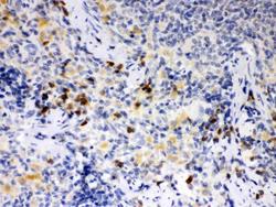
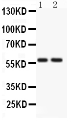
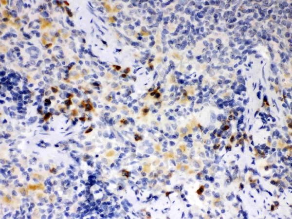
- Main image
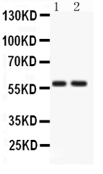
- Experimental details
- Western blot analysis of ATG14L using anti-ATG14L antibody (PB9481). Electrophoresis was performed on a 5-20% SDS-PAGE gel at 70V (Stacking gel) / 90V (Resolving gel) for 2-3 hours. The sample well of each lane was loaded with 50ug of sample under reducing conditions. Lane 1: Rat Brain Tissue Lysate, Lane 2: HELA Whole Cell Lysate. After Electrophoresis, proteins were transferred to a Nitrocellulose membrane at 150mA for 50-90 minutes. Blocked the membrane with 5% Non-fat Milk/ TBS for 1.5 hour at RT. The membrane was incubated with rabbit anti-ATG14L antigen affinity purified polyclonal antibody (Catalog # PB9481) at 0.5 μg/mL overnight at 4°C, then washed with TBS-0.1%Tween 3 times with 5 minutes each and probed with a goat anti-rabbit IgG-HRP secondary antibody at a dilution of 1:10000 for 1.5 hour at RT. The signal is developed using an Enhanced Chemiluminescent detection (ECL) kit (Catalog # EK1002) with Tanon 5200 system. A specific band was detected for ATG14L at approximately 59KD. The expected band size for ATG14L is at 59KD.
- Additional image