 Explore
 Validate
 Learn
Learn Western blot
 Immunocytochemistry
 Immunohistochemistry
Antibody data
- Antibody Data
- Antigen structure
- References [1]
- Comments [0]
- Validations
- Immunocytochemistry [2]
- Other assay [1]
Submit
Validation data
Reference
Comment
Report error
- Product number
- PA1-084 - Provider product page

- Provider
- Invitrogen Antibodies
- Product name
- RANBP3 Polyclonal Antibody
- Antibody type
- Polyclonal
- Antigen
- Synthetic peptide
- Description
- PA1-084 detects Ran binding protein 3 (RanBP3) from human and rat cells. PA1-084 has been successfully used in Western blot and immunofluorescence procedures. By Western blot this antibody detects an ~60 kDa protein representing RanBP3 from HeLa cell extracts. Immunofluorescence staining of RanBP3 in NRK cells with PA1-084 results in diffuse nuclear staining. The PA1-084 immunogen is a synthetic peptide corresponding to residues M(1) A D L A N E E K P A L A(13) C of human RanBP3. The PA1-084 immunizing peptide (Cat. # PEP-177) is available for use in neutralization and control experiments.
- Reactivity
- Human, Rat
- Host
- Rabbit
- Isotype
- IgG
- Vial size
- 200 μg
- Concentration
- 1 mg/mL
- Storage
- -20°C, Avoid Freeze/Thaw Cycles
Submitted references Sphingosine kinase 1 serves as a pro-viral factor by regulating viral RNA synthesis and nuclear export of viral ribonucleoprotein complex upon influenza virus infection.
Seo YJ, Pritzl CJ, Vijayan M, Bomb K, McClain ME, Alexander S, Hahm B
PloS one 2013;8(8):e75005
PloS one 2013;8(8):e75005
No comments: Submit comment
Supportive validation
- Submitted by
- Invitrogen Antibodies (provider)
- Main image

- Experimental details
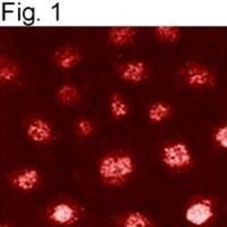
- Immunocytochemical staining of RanBP3 in NRK cells using Product # PA1-084.

- Submitted by
- Invitrogen Antibodies (provider)
- Main image
- Experimental details
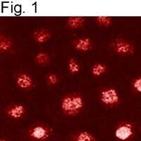
- Immunocytochemical staining of RanBP3 in NRK cells using Product # PA1-084.
Supportive validation
- Submitted by
- Invitrogen Antibodies (provider)
- Main image
- Experimental details
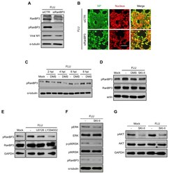
- Figure 5 SK inhibition impairs virus-induced activation of ERK/p90RSK/AKT to inhibit RanBP3-mediated nuclear export of viral NP. (A and B) HEK293 cells were transfected with scramble siRNA (siCTR) or siRNA targeting RanBP3 (siRanBP3); then the cells were infected with influenza virus at an MOI of 0.01 (A) or 1 (B). RanBP3, pRanBP3, viral M1, and alpha-tubulin were detected by Western blotting at 30 hpi (A). Viral NP (green) and nuclei (DRAQ5 dye, red) were visualized by confocal microscopy at 12 hpi (B). (C and D) MDCK cells were treated with solvent, DMS (5 uM), or SKI-II (10 uM) and infected with influenza virus at an MOI of 5 for the indicated times (C) or 6 hrs (D). Cell lysates were used for Western blot analysis to detect pRanBP3, RanBP3, alpha-tubulin, or actin. (E) MDCK cells were left untreated or treated with U0126 (10 uM) or LY294002 (10 uM) and infected with influenza virus at 3 MOI for 6 hrs. Western blot analysis was performed to detect pRanBP3, RanBP3, and GAPDH. (F) MDCK cells were treated with solvent alone or SKI-II (10 uM) and infected with influenza virus at an MOI of 1. Western blot analysis was performed to detect pERK, ERK, p-p90RSK, p90RSK, pRANBP3, or alpha-tubulin at 12 hpi. (G) MDCK cells were treated with solvent or SKI-II (10 uM) and mock-infected or infected with influenza virus at an MOI of 3. At 8 hpi, pAKT, AKT, and alpha-tubulin were detected by Western blot analysis.
