Explore
 Validate
 Learn
LearnHPA008999
antibody from Atlas Antibodies
Targeting: EFNB2
EPLG5, Htk-L, HTKL, LERK5, MGC126226, MGC126227, MGC126228
 Western blot
 Immunocytochemistry
Antibody data
- Antibody Data
- Antigen structure
- References [8]
- Comments [0]
- Validations
- Immunocytochemistry [1]
Submit
Validation data
Reference
Comment
Report error
- Product number
- HPA008999 - Provider product page

- Provider
- Atlas Antibodies
- Proper citation
- Atlas Antibodies Cat#HPA008999, RRID:AB_1078721
- Product name
- Anti-EFNB2
- Antibody type
- Polyclonal
- Description
- Polyclonal Antibody against Human EFNB2, Gene description: ephrin-B2, Alternative Gene Names: EPLG5, Htk-L, HTKL, LERK5, MGC126226, MGC126227, MGC126228, Validated applications: ICC, IHC, WB, Uniprot ID: P52799, Storage: Store at +4°C for short term storage. Long time storage is recommended at -20°C.
- Reactivity
- Human
- Host
- Rabbit
- Conjugate
- Unconjugated
- Isotype
- IgG
- Vial size
- 100 µl
- Concentration
- 0.1 mg/ml
- Storage
- Store at +4°C for short term storage. Long time storage is recommended at -20°C.
- Handling
- The antibody solution should be gently mixed before use.
Submitted references Activation of EphrinB2/EphB2 signaling in the spine cord alters glia-neuron interactions in mice with visceral hyperalgesia following maternal separation
M2 macrophage-derived cathepsin S promotes peripheral nerve regeneration via fibroblast–Schwann cell-signaling relay
Placenta Percreta Presents with Neoangiogenesis of Arteries with Von Willebrand Factor-Negative Endothelium
Dysregulation of the EphrinB2−EphB4 ratio in pediatric cerebral arteriovenous malformations is associated with endothelial cell dysfunction in vitro and functions as a novel noninvasive biomarker in patients
ADAM10-mediated ephrin-B2 shedding promotes myofibroblast activation and organ fibrosis
Interrogating cellular fate decisions with high-throughput arrays of multiplexed cellular communities
Ephrin-B2 controls PDGFRβ internalization and signaling
Spatial regulation of VEGF receptor endocytosis in angiogenesis
Guo S, Wang Y, Duan Q, Gu W, Fu Q, Ma Z, Ruan J
Frontiers in Pharmacology 2024;15
Frontiers in Pharmacology 2024;15
M2 macrophage-derived cathepsin S promotes peripheral nerve regeneration via fibroblast–Schwann cell-signaling relay
Oshima E, Hayashi Y, Xie Z, Sato H, Hitomi S, Shibuta I, Urata K, Ni J, Iwata K, Shirota T, Shinoda M
Journal of Neuroinflammation 2023;20(1)
Journal of Neuroinflammation 2023;20(1)
Placenta Percreta Presents with Neoangiogenesis of Arteries with Von Willebrand Factor-Negative Endothelium
Schwickert A, Henrich W, Vogel M, Melchior K, Ehrlich L, Ochs M, Braun T
Reproductive Sciences 2021;29(4):1136-1144
Reproductive Sciences 2021;29(4):1136-1144
Dysregulation of the EphrinB2−EphB4 ratio in pediatric cerebral arteriovenous malformations is associated with endothelial cell dysfunction in vitro and functions as a novel noninvasive biomarker in patients
Fehnel K, Penn D, Duggins-Warf M, Gruber M, Pineda S, Sesen J, Moses-Gardner A, Shah N, Driscoll J, Zurakowski D, Orbach D, Smith E
Experimental & Molecular Medicine 2020;52(4):658-671
Experimental & Molecular Medicine 2020;52(4):658-671
ADAM10-mediated ephrin-B2 shedding promotes myofibroblast activation and organ fibrosis
Lagares D, Ghassemi-Kakroodi P, Tremblay C, Santos A, Probst C, Franklin A, Santos D, Grasberger P, Ahluwalia N, Montesi S, Shea B, Black K, Knipe R, Blati M, Baron M, Wu B, Fahmi H, Gandhi R, Pardo A, Selman M, Wu J, Pelletier J, Martel-Pelletier J, Tager A, Kapoor M
Nature Medicine 2017;23(12):1405-1415
Nature Medicine 2017;23(12):1405-1415
Interrogating cellular fate decisions with high-throughput arrays of multiplexed cellular communities
Chen S, Bremer A, Scheideler O, Na Y, Todhunter M, Hsiao S, Bomdica P, Maharbiz M, Gartner Z, Schaffer D
Nature Communications 2016;7(1)
Nature Communications 2016;7(1)
Ephrin-B2 controls PDGFRβ internalization and signaling
Nakayama A, Nakayama M, Turner C, Höing S, Lepore J, Adams R
Genes & Development 2013;27(23):2576-2589
Genes & Development 2013;27(23):2576-2589
Spatial regulation of VEGF receptor endocytosis in angiogenesis
Nakayama M, Nakayama A, van Lessen M, Yamamoto H, Hoffmann S, Drexler H, Itoh N, Hirose T, Breier G, Vestweber D, Cooper J, Ohno S, Kaibuchi K, Adams R
Nature Cell Biology 2013;15(3):249-260
Nature Cell Biology 2013;15(3):249-260
No comments: Submit comment
Supportive validation
- Submitted by
- Atlas Antibodies (provider)
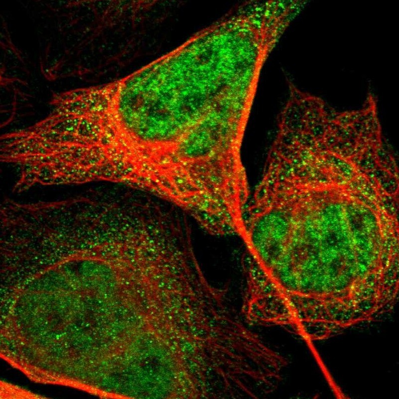
- Main image

- Experimental details
- Immunofluorescent staining of human cell line U-2 OS shows localization to nucleoplasm & cytosol.
- Sample type
- Human