Explore
 Validate
 Learn
Learn Western blot
 ELISA
ELISAAntibody data
- Antibody Data
- Antigen structure
- References [0]
- Comments [0]
- Validations
- Western blot [4]
- Immunohistochemistry [1]
- Flow cytometry [1]
Submit
Validation data
Reference
Comment
Report error
- Product number
- NBP1-04261 - Provider product page

- Provider
- Novus Biologicals
- Proper citation
- Novus Cat#NBP1-04261, RRID:AB_1520204
- Product name
- Mouse Monoclonal AK3 Antibody
- Antibody type
- Monoclonal
- Description
- Protein G purified.
- Reactivity
- Human
- Host
- Mouse
- Isotype
- IgG
- Vial size
- 0.1 ml
- Concentration
- 1.0 mg/ml
- Storage
- Store at 4C short term. Aliquot and store at -20C long term. Avoid freeze-thaw cycles.
No comments: Submit comment
Supportive validation
- Submitted by
- Novus Biologicals (provider)
- Main image
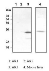
- Experimental details
- Western Blot: AK3 Antibody (SJB3-36) [NBP1-04261] - The recombinant human AK isozymes (AK1, AK2, AK3) and mouse liver were resolved by SDS-PAGE, transferred to PVDF membrane and probed with anti-AK3 antibody (1:1,000). Proteins were visualized using a goat anti-mouse secondary antibody conjugated to HRP.
- Submitted by
- Novus Biologicals (provider)
- Main image
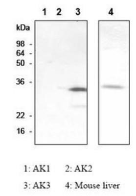
- Experimental details
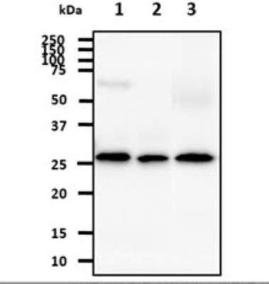
- Western Blot: AK3 Antibody (SJB3-36) [NBP1-04261] - The recombinant protein (50ng) were resolved by SDS-PAGE, transferred to PVDF membrane and probed with anti-human AK3 antibody (1:1,000). Proteins were visualized using a goat anti-mouse secondary antibody conjugated to HRP and an ECL detection system. Lane 1. : Recombinant Human AK1 Lane 2. : Recombinant Human AK2 Lane 3. : Recombinant Human AK3

- Submitted by
- Novus Biologicals (provider)
- Main image

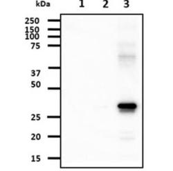
- Experimental details
- Western Blot: AK3 Antibody (SJB3-36) [NBP1-04261] - The lysate (40ug) were resolved by SDS-PAGE, transferred to NC membrane and probed with anti-human AK3 antibody (1:1000). Proteins were visualized using a goat anti-mouse secondary antibody conjugated to HRP and an ECL detection system. Lane 1. : Hep3B cell lysate Lane 2. : 293T cell lysate Lane 3. : Mouse kidney tissue lysate
- Submitted by
- Novus Biologicals (provider)
- Main image

- Experimental details
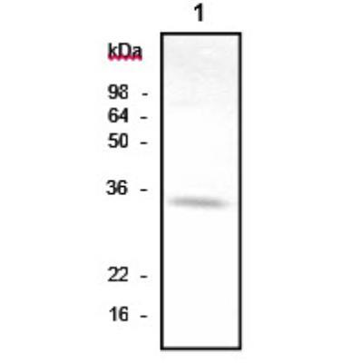
- Western Blot: AK3 Antibody (SJB3-36) [NBP1-04261] - The lysate (40ug) were resolved by SDS-PAGE, transferred to NC membrane and probed with anti-human AK3 antibody (1:1000). Proteins were visualized using a goat anti-mouse secondary antibody conjugated to HRP and an ECL detection system. Lane 1. : Mouse liver tissue lysate
Supportive validation
- Submitted by
- Novus Biologicals (provider)
- Main image
- Experimental details
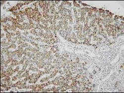
- Immunohistochemistry-Paraffin: AK3 Antibody (SJB3-36) [NBP1-04261] - Human liver tissue was incubated with anti-human AK3 (1:100) for 2 hours at room temperature. Slide was then washed in PBS, and was incubated in avidin biosystem anti-rabbit labeled polymer for 30 min at RT. Enzyme detection was performed with DAB chromo-gen.

Supportive validation
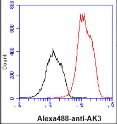
- Submitted by
- Novus Biologicals (provider)
- Main image
- Experimental details
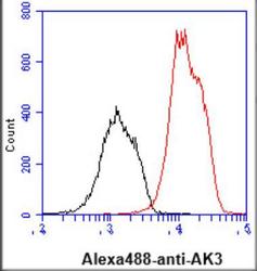
- Flow Cytometry: AK3 Antibody (SJB3-36) [NBP1-04261] - Analysis of AK3 in jurkat cell line, staining at 2-5ug for 1x10⁶cells (red line). The secondary antibody used goat anti-mouse IgG Alexa fluor 488 conjugate. Isotype control antibody was mouse IgG (black line).