Explore
 Validate
 Learn
Learn Western blot
 Immunoprecipitation
Antibody data
- Antibody Data
- Antigen structure
- References [0]
- Comments [0]
- Validations
- Western blot [2]
- Immunocytochemistry [1]
- Other assay [1]
Submit
Validation data
Reference
Comment
Report error
- Product number
- 710012 - Provider product page

- Provider
- Invitrogen Antibodies
- Product name
- Anti-CENTB1 Recombinant Polyclonal Antibody (19HCLC)
- Antibody type
- Other
- Antigen
- Other
- Description
- This antibody is predicted to react with mouse based on sequence homology. Recombinant rabbit polyclonal antibodies are unique offerings from Thermo Fisher Scientific. They are comprised of a selection of multiple different recombinant monoclonal antibodies, providing the best of both worlds - the sensitivity of polyclonal antibodies with the specificity of monoclonal antibodies - all delivered with the consistency only found in a recombinant antibody. While functionally the same as a polyclonal antibody - recognizing multiple epitope sites on the target and producing higher detection sensitivity for low abundance targets - a recombinant rabbit polyclonal antibody has a known mixture of light and heavy chains. The exact population can be produced in every lot, circumventing the biological variability typically associated with polyclonal antibody production.
- Reactivity
- Human
- Host
- Rabbit
- Isotype
- IgG
- Antibody clone number
- 19HCLC
- Vial size
- 100 µg
- Concentration
- 0.5 mg/mL
- Storage
- Store at 4°C short term. For long term storage, store at -20°C, avoiding freeze/thaw cycles.
No comments: Submit comment
Supportive validation
- Submitted by
- Invitrogen Antibodies (provider)
- Main image
- Experimental details
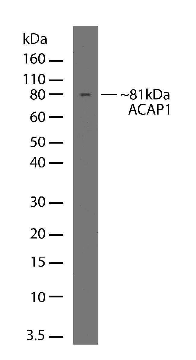
- Western blot analysis was performed on whole cell extracts (50 µg lysate) of Jurkat. The blots were probed with Anti-ACAP1 Recombinant Rabbit Polyclonal Antibody (Product # 710012, 4-6 µg/mL) and detected by chemiluminescence Goat anti-Rabbit IgG (H+L) Superclonal™ Secondary Antibody, HRP conjugate (Product # A27036, 0.4 µg/mL, 1:2500 dilution). A 81 kDa band corresponding to ACAP1 was observed. Known quantity of protein samples were electrophoresed using Novex® NuPAGE® 12 % Bis-Tris gel (Product # NP0342BOX), XCell SureLock™ Electrophoresis System (Product # EI0002) and Novex® Sharp Pre-Stained Protein Standard (Product # LC5800). Resolved proteins were then transferred onto a nitrocellulose membrane with iBlot® 2 Dry Blotting System (Product # IB21001). The membrane was probed with the relevant primary and secondary Antibody following blocking with 5 % skimmed milk. Chemiluminescent detection was performed using Pierce™ ECL Western blotting Substrate (Product # 32106).

- Submitted by
- Invitrogen Antibodies (provider)
- Main image

- Experimental details
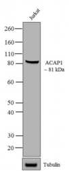
- Western blot analysis of ACAP1 in Jurkat cell lysate (30 µg/lane) using an ACAP1 Recombinant Rabbit Polyclonal Antibody (Product # 710012) at a dilution of 5 µg/mL. NBT/BCIP was used as the substrate (Product # WB7105).
Supportive validation
- Submitted by
- Invitrogen Antibodies (provider)
- Main image
- Experimental details
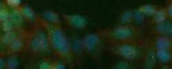
- Immunofluorescent analysis of CENTB1 in HeLa cells using a CENTB1 Recombinant Rabbit Polyclonal Antibody (Product # 710012) at a dilution of 10 µg/mL followed by detection using an Alexa Fluor 488-conjugated Goat anti-Rabbit secondary antibody at a dilution of 1:1000 (green), nuclei staining using DAPI (blue) and Alexa Fluor 594 Phalloidin (red). The cellular localization is predominantly cytoplasmic.

Supportive validation
- Submitted by
- Invitrogen Antibodies (provider)
- Main image

- Experimental details
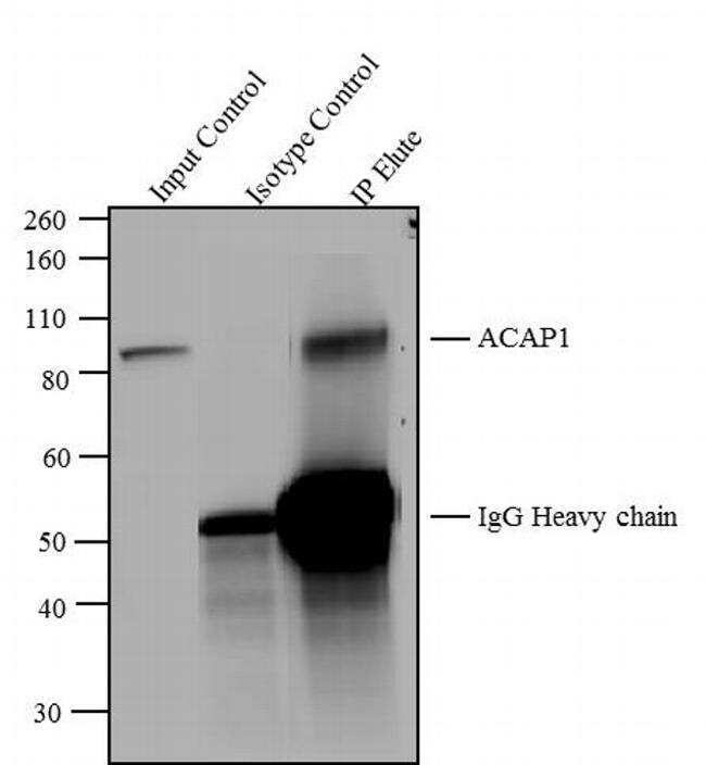
- ACAP1 was immunoprecipitated using 5 µg of the ACAP1 Recombinant Rabbit Polyclonal Antibody (Product # 710012) from lysate of Jurkat cells (Lane 3) using the Dynabeads® Protein A Immunoprecipitation Kit (Product #10006D). Normal Rabbit IgG was used as a Isotype control (Lane 2). 10 % input represents the cell extract used for immunoprecipitation (Lane 1). Western blot analysis was performed using ACAP1 Recombinant Rabbit Polyclonal Antibody (Product # 710012) and Goat anti-Rabbit IgG (H+L) Superclonal™ Secondary Antibody, HRP conjugate (Product # A27036, 0.4 µg/mL, 1:2500 dilution). Chemiluminescent detection was performed using Pierce™ ECL Western blotting Substrate (Product # 32106).