 Explore
 Validate
 Learn
Learn Western blot
Antibody data
- Antibody Data
- Antigen structure
- References [0]
- Comments [0]
- Validations
- Western blot [2]
- Immunohistochemistry [1]
Submit
Validation data
Reference
Comment
Report error
- Product number
- ANC-020-200UL - Provider product page

- Provider
- Invitrogen Antibodies
- Product name
- RIC3 Polyclonal Antibody
- Antibody type
- Polyclonal
- Antigen
- Other
- Reactivity
- Human, Mouse, Rat
- Host
- Rabbit
- Isotype
- IgG
- Vial size
- 200 µL
- Concentration
- 0.8 mg/mL
- Storage
- -20° C, Avoid Freeze/Thaw Cycles
No comments: Submit comment
Supportive validation
- Submitted by
- Invitrogen Antibodies (provider)
- Main image
- Experimental details
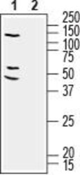
- Western blot analysis of human SH-SY5Y neuroblastoma cell line lysate: - 1. Anti-RIC3 Antibody (#ANC-020), (1:200). 2. Anti-RIC3 Antibody , preincubated with RIC3 Blocking Peptide (#BLP-NC020).
- Submitted by
- Invitrogen Antibodies (provider)
- Main image
- Experimental details
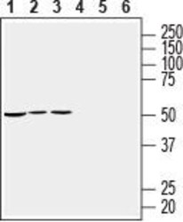
- Western blot analysis of mouse heart membranes (lanes 1 and 4), rat heart lysate (lanes 2 and 5) and rat brain lysate (lanes 3 and 6): - 1-3. Anti-RIC3 Antibody (#ANC-020), (1:200).4-6. Anti-RIC3 Antibody , preincubated with RIC3 Blocking Peptide (#BLP-NC020).

Supportive validation
- Submitted by
- Invitrogen Antibodies (provider)
- Main image
- Experimental details
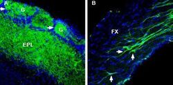
- Expression of RIC-3 in mouseolfactory bulbandfornix - Immunohistochemical staining of perfusion-fixed frozen mouse brain sections with Anti-RIC3 Antibody (#ANC-020), (1:400), followed by goat- Anti-rabbit-AlexaFluor-488. A. RIC-3 staining in mouse olfactory bulb shows staining in the external plexiform layer (EPL) and in some glomeruli (G). Arrows point at glomeruli with RIC-3 positive core. B. RIC-3 staining in mouse fornix is detected in neuronal profiles (vertical arrows) and neuronal processes (horizontal arrow). Cell nuclei are stained with DAPI (blue).
