Explore
 Validate
 Learn
Learn12-0938-41
antibody from Invitrogen Antibodies
Targeting: CD93
C1qR(P), C1QR1, C1qRP, CDw93, dJ737E23.1, ECSM3, MXRA4
 Flow cytometry
Antibody data
- Antibody Data
- Antigen structure
- References [3]
- Comments [0]
- Validations
- Flow cytometry [1]
Submit
Validation data
Reference
Comment
Report error
- Product number
- 12-0938-41 - Provider product page

- Provider
- Invitrogen Antibodies
- Product name
- Anti-CD93 (AA4.1) Monoclonal Antibody (R3), PE, eBioscience™
- Antibody type
- Monoclonal
- Antigen
- Other
- Description
- Description: The monoclonal antibody R3 recognizes human CD93, also known as C1qRp. The glycoprotein CD93 binds to C1q, the subunit of the complement protein, mannose binding lectin and pulmonary surfactant protein A. CD93 is predicted to play a role in the clearance of apoptotic cells. Expression of CD93 is confined to myeloid cells with higher expression on monocytes than neutrophils, eosinophils, platelets and endothelial cells. Expression on DC's is downregulated upon maturation. Additionally, CD93 has been shown to define an early bone marrow stem cell population of hematopoietic and hepatic precursors. The R3 antibody has been shown to have activating function by increasing phagocytosis when presented in an immobilized state and inhibitory functions when presented in a soluble state. CD93 can be shed from the cell surface. This phenomenon can be measures using R3 antibody as detection with R139 as capture to detect soluble CD93 by ELISA. The epitope for R3 lies in the CRD domain.\\The R3 antibody has been shown to have activating function by increasing phagocytosis when presented in an immobilized state and inhibitory functions when presented in a soluble state. CD93 can be shed from the cell surface. This phenomenon can be measures using R3 antibody as detection with R139 as capture to detect soluble CD93 by ELISA. The epitope for R3 lies in the CRD domain. Applications Reported: This R3 antibody has been reported for use in flow cytometric analysis. Applications Tested: This R3 antibody has been pre-titrated and tested by flow cytometric analysis of normal human peripheral blood cells. This can be used at 5 µL (0.125 µg) per test. A test is defined as the amount (µg) of antibody that will stain a cell sample in a final volume of 100 µL. Cell number should be determined empirically but can range from 10^5 to 10^8 cells/test. Excitation: 488-561 nm; Emission: 578 nm; Laser: Blue Laser, Green Laser, Yellow-Green Laser. Filtration: 0.2 µm post-manufacturing filtered.
- Reactivity
- Human
- Host
- Mouse
- Conjugate
- Yellow dye
- Isotype
- IgM
- Antibody clone number
- R3
- Vial size
- 25 Tests
- Concentration
- 5 µL/Test
- Storage
- 4° C, store in dark, DO NOT FREEZE!
Submitted references CD93 is rapidly shed from the surface of human myeloid cells and the soluble form is detected in human plasma.
C1qRp defines a new human stem cell population with hematopoietic and hepatic potential.
C1qRP, the C1q receptor that enhances phagocytosis, is detected specifically in human cells of myeloid lineage, endothelial cells, and platelets.
Bohlson SS, Silva R, Fonseca MI, Tenner AJ
Journal of immunology (Baltimore, Md. : 1950) 2005 Jul 15;175(2):1239-47
Journal of immunology (Baltimore, Md. : 1950) 2005 Jul 15;175(2):1239-47
C1qRp defines a new human stem cell population with hematopoietic and hepatic potential.
Danet GH, Luongo JL, Butler G, Lu MM, Tenner AJ, Simon MC, Bonnet DA
Proceedings of the National Academy of Sciences of the United States of America 2002 Aug 6;99(16):10441-5
Proceedings of the National Academy of Sciences of the United States of America 2002 Aug 6;99(16):10441-5
C1qRP, the C1q receptor that enhances phagocytosis, is detected specifically in human cells of myeloid lineage, endothelial cells, and platelets.
Nepomuceno RR, Tenner AJ
Journal of immunology (Baltimore, Md. : 1950) 1998 Feb 15;160(4):1929-35
Journal of immunology (Baltimore, Md. : 1950) 1998 Feb 15;160(4):1929-35
No comments: Submit comment
Supportive validation
- Submitted by
- Invitrogen Antibodies (provider)
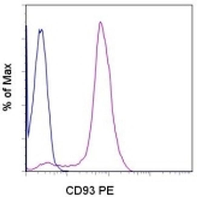
- Main image
- Experimental details
- Staining of normal human peripheral blood cells with Mouse IgM Isotype Control PE (blue histogram) or Anti-Human CD93 PE (purple histogram). Cells in the monocyte gate were used for analysis.
- Conjugate
- Yellow dye