Explore
 Validate
 Learn
Learn Western blot
 Immunocytochemistry
Antibody data
- Antibody Data
- Antigen structure
- References [3]
- Comments [0]
- Validations
- Immunocytochemistry [2]
- Other assay [2]
Submit
Validation data
Reference
Comment
Report error
- Product number
- 702434 - Provider product page

- Provider
- Invitrogen Antibodies
- Product name
- JAK2 Recombinant Rabbit Monoclonal Antibody (18H11L8)
- Antibody type
- Monoclonal
- Antigen
- Synthetic peptide
- Description
- This antibody is predicted to react with Monkey, Rat, Sheep and Bovine. Recombinant rabbit monoclonal antibodies are produced using in vitro expression systems. The expression systems are developed by cloning in the specific antibody DNA sequences from immunoreactive rabbits. Then, individual clones are screened to select the best candidates for production. The advantages of using recombinant rabbit monoclonal antibodies include: better specificity and sensitivity, lot-to-lot consistency, animal origin-free formulations, and broader immunoreactivity to diverse targets due to larger rabbit immune repertoire.
- Reactivity
- Human
- Host
- Rabbit
- Isotype
- IgG
- Antibody clone number
- 18H11L8
- Vial size
- 100 μg
- Concentration
- 0.5 mg/mL
- Storage
- Store at 4°C short term. For long term storage, store at -20°C, avoiding freeze/thaw cycles.
Submitted references Discovery of a novel potentially transforming somatic mutation in CSF2RB gene in breast cancer.
Isolation and Establishment of a Highly Proliferative, Cancer Stem Cell-Like, and Naturally Immortalized Triple-Negative Breast Cancer Cell Line, KAIMRC2.
Role and mechanism of nursing cooperation and tetramethylpyrazine application in post-operative pain in patients undergoing total knee arthroplasty.
Rashid M, Ali R, Almuzzaini B, Song H, AlHallaj A, Abdulkarim AA, Mohamed Baz O, Al Zahrani H, Mustafa Sabeena M, Alharbi W, Hussein M, Boudjelal M
Cancer medicine 2021 Nov;10(22):8138-8150
Cancer medicine 2021 Nov;10(22):8138-8150
Isolation and Establishment of a Highly Proliferative, Cancer Stem Cell-Like, and Naturally Immortalized Triple-Negative Breast Cancer Cell Line, KAIMRC2.
Ali R, Al Zahrani H, Barhoumi T, Alhallaj A, Mashhour A, Alshammari MA, Alshawakir YA, Baz O, Alanazi AH, Khan AL, Al Nikhli H, Al Balwi MA, Al Riyees L, Boudjelal M
Cells 2021 May 24;10(6)
Cells 2021 May 24;10(6)
Role and mechanism of nursing cooperation and tetramethylpyrazine application in post-operative pain in patients undergoing total knee arthroplasty.
Liu C, Liu R, Tang M, Yang X, Gong X
Experimental and therapeutic medicine 2019 Mar;17(3):2366-2372
Experimental and therapeutic medicine 2019 Mar;17(3):2366-2372
No comments: Submit comment
Supportive validation
- Submitted by
- Invitrogen Antibodies (provider)
- Main image
- Experimental details
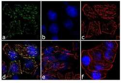
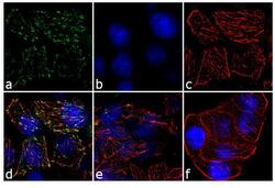
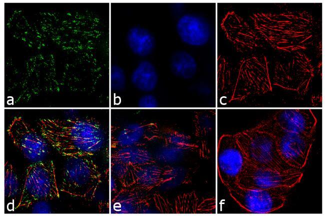
- For immunofluorescence analysis, MG-132 (20 uM, 1 h) and IFN gamma (100 ng, 15 min) treated HT-29 cells were fixed and permeabilized for detection of endogenous Jak2 using Anti-Jak2 Recombinant Rabbit Monoclonal Antibody (Product # 702434, 2 µg/mL) and labeled with Goat anti-Rabbit IgG (H+L) Superclonal™ Secondary Antibody, Alexa Fluor® 488 conjugate (Product # A27034, 1:2000). Panel a) shows representative cells that were stained for detection and localization of Jak2 protein (green), Panel b) is stained for nuclei (blue) using SlowFade® Gold Antifade Mountant with DAPI (Product # S36938). Panel c) represents cytoskeletal F-actin staining using Alexa Fluor® 555 Rhodamine Phalloidin (Product # R415, 1:300). Panel d) is a composite image of Panels a, b and c clearly demonstrating membrane localization of Jak2. Panel e) shows untreated cells with no signal. Panel f) represents control cells with no primary antibody to assess background. The images were captured at 60X magnification.
- Submitted by
- Invitrogen Antibodies (provider)
- Main image
- Experimental details
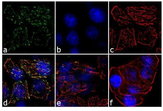
- For immunofluorescence analysis, MG-132 (20 uM, 1 h) and IFN gamma (100 ng, 15 min) treated HT-29 cells were fixed and permeabilized for detection of endogenous Jak2 using Anti-Jak2 Recombinant Rabbit Monoclonal Antibody (Product # 702434, 2 µg/mL) and labeled with Goat anti-Rabbit IgG (Heavy Chain) Superclonal™ Secondary Antibody, Alexa Fluor® 488 conjugate (Product # A27034, 1:2000). Panel a) shows representative cells that were stained for detection and localization of Jak2 protein (green), Panel b) is stained for nuclei (blue) using SlowFade® Gold Antifade Mountant with DAPI (Product # S36938). Panel c) represents cytoskeletal F-actin staining using Alexa Fluor® 555 Rhodamine Phalloidin (Product # R415, 1:300). Panel d) is a composite image of Panels a, b and c clearly demonstrating membrane localization of Jak2. Panel e) shows untreated cells with no signal. Panel f) represents control cells with no primary antibody to assess background. The images were captured at 60X magnification.
Supportive validation
- Submitted by
- Invitrogen Antibodies (provider)
- Main image
- Experimental details
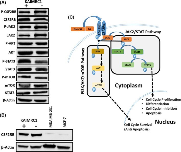
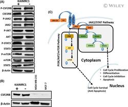
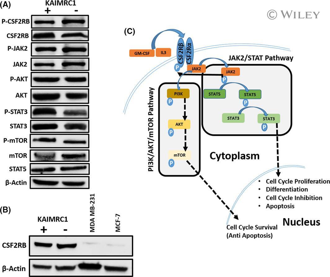
- 2 FIGURE Characterization of KAIMRC1 cells on protein level. (A) Western blot analysis of the KAIMRC1 cell line in normal (+) and serum-starved conditions(-) against P- CSF2RB , CSF2RB , P-AKT1, AKT1, JAK2, P-JAK2, STAT3, P-STAT3, STAT5, mTOR, and P-mTOR. Ligand-independent activation of AKT/mTOR pathway, as well as the JAK2/STAT pathway, was observed. (B) Comparative Western blot analysis of KAIMRC1, MDA-MB-231, and MCF-7 cell lines against CSF2RB . (C) Schematic representation of proposed activation of the AKT/mTOR pathway and JAK2/STAT pathway that might result in cell cycle survival and proliferation
- Submitted by
- Invitrogen Antibodies (provider)
- Main image
- Experimental details
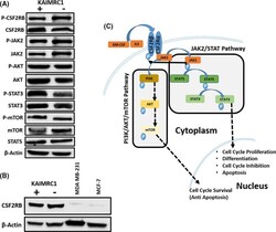
- FIGURE 2 Characterization of KAIMRC1 cells on protein level. (A) Western blot analysis of the KAIMRC1 cell line in normal (+) and serum-starved conditions(-) against P- CSF2RB , CSF2RB , P-AKT1, AKT1, JAK2, P-JAK2, STAT3, P-STAT3, STAT5, mTOR, and P-mTOR. Ligand-independent activation of AKT/mTOR pathway, as well as the JAK2/STAT pathway, was observed. (B) Comparative Western blot analysis of KAIMRC1, MDA-MB-231, and MCF-7 cell lines against CSF2RB . (C) Schematic representation of proposed activation of the AKT/mTOR pathway and JAK2/STAT pathway that might result in cell cycle survival and proliferation