Explore
 Validate
 Learn
Learn Western blot
Antibody data
- Antibody Data
- Antigen structure
- References [0]
- Comments [0]
- Validations
- Western blot [3]
- Immunocytochemistry [5]
- Immunohistochemistry [1]
Submit
Validation data
Reference
Comment
Report error
- Product number
- PA5-21392 - Provider product page

- Provider
- Invitrogen Antibodies
- Product name
- RRBP1 Polyclonal Antibody
- Antibody type
- Polyclonal
- Antigen
- Recombinant protein fragment
- Description
- Recommended positive controls: HepG2, NIH-3T3. Predicted reactivity: Mouse (81%), Dog (83%). Store product as a concentrated solution. Centrifuge briefly prior to opening the vial.
- Reactivity
- Human, Mouse
- Host
- Rabbit
- Isotype
- IgG
- Vial size
- 100 µL
- Concentration
- 1 mg/mL
- Storage
- Store at 4°C short term. For long term storage, store at -20°C, avoiding freeze/thaw cycles.
No comments: Submit comment
Supportive validation
- Submitted by
- Invitrogen Antibodies (provider)
- Main image

- Experimental details
- Western Blot analysis of RRBP1 was performed by separating various whole cell lysates by 5% SDS-PAGE. Proteins were transferred to a membrane and probed with a RRBP1 Polyclonal Antibody (Product # PA5-21392) at a dilution of 1:1000. The HRP-conjugated anti-rabbit IgG antibody was used to detect the primary antibody. Upperpanel: Ribosome binding protein 1 antibody, Lower panel: Vinculin antibody, B. 30 µg of RRBP1 siRNA1-transfected HepG2 cells, C. 30 µg of RRBP1 siRNA2-transfected HepG2 cells.

- Submitted by
- Invitrogen Antibodies (provider)
- Main image

- Experimental details
- Western Blot using RRBP1 Polyclonal Antibody (Product # PA5-21392). Sample (30 µg of whole cell lysate). Lane A: NIH-3T3.5% SDS PAGE. RRBP1 Polyclonal Antibody (Product # PA5-21392) diluted at 1:1,000. The HRP-conjugated anti-rabbit IgG antibody was used to detect the primary antibody.

- Submitted by
- Invitrogen Antibodies (provider)
- Main image

- Experimental details
- Western blot analysis was performed on whole cell extract (30 µg lysate) of MKN45 (Lane 1), HCT116 (Lane 2), GTL16 (Lane 3), HepG2 (Lane 4), A549 (Lane 5), K562 (Lane 6), HeLa (Lane 7), Raji (Lane 8) and L6 (Lane 9). The blot was probed with Anti-RRBP1 Polyclonal Antibody (Product # PA5-21392, 1:8000 dilution) and detected by chemiluminescence using Goat anti-Rabbit IgG (H+L) Superclonal™ Secondary Antibody, HRP conjugate (Product # A27036, 0.25 µg/ml, 1:4000 dilution). Bands of ~152 and ~108 kDa corresponding to two different isoforms of RRBP1 was observed in cell lines tested.

Supportive validation
- Submitted by
- Invitrogen Antibodies (provider)
- Main image

- Experimental details
- Immunofluorescent analysis of ribosome binding protein 1 in methanol-fixed HeLa cells using a ribosome binding protein 1 polyclonal antibody (Product # PA5-21392) (Green) at a 1:500 dilution. Alpha-tubulin filaments were labeled with Product # PA5-29281 (Red) at a 1:2500.

- Submitted by
- Invitrogen Antibodies (provider)
- Main image

- Experimental details
- Immunofluorescent analysis of ribosome binding protein 1 in paraformaldehyde-fixed HeLa cells using a ribosome binding protein 1 polyclonal antibody (Product # PA5-21392) (Green) at a 1:500 dilution. Alpha-tubulin filaments were labeled with Product # PA5-29281 (Red) at a 1:500.

- Submitted by
- Invitrogen Antibodies (provider)
- Main image

- Experimental details
- Confocal immunofluorescence analysis (Olympus FV10i) of paraformaldehyde-fixed HeLa, using Ribosome binding protein 1 antibody (Product # PA5-21392) (Green) at 1:500 dilution. Alpha-tubulin filaments were labeled with (Product # MA1-25054) (Red) at 1:500.

- Submitted by
- Invitrogen Antibodies (provider)
- Main image

- Experimental details
- Confocal immunofluorescence analysis (Olympus FV10i) of methanol-fixed HeLa, using Ribosome binding protein 1 antibody (Product # PA5-21392) (Green) at 1:500 dilution. Alpha-tubulin filaments were labeled with (Product # MA1-25054) (Red) at 1:2500.

- Submitted by
- Invitrogen Antibodies (provider)
- Main image

- Experimental details
- Immunofluorescence analysis of RRBP1 was performed using 70% confluent log phase A549 cells. The cells were fixed with 4% paraformaldehyde for 10 minutes, permeabilized with 0.1% Triton™ X-100 for 15 minutes, and blocked with 1% BSA for 1 hour at room temperature. The cells were labeled with RRBP1 Polyclonal Antibody (Product # PA5-21392) at 1:250 dilution in 0.1% BSA, incubated at 4 degree Celsius overnight and then labeled with Goat anti-Rabbit IgG (H+L) Superclonal™ Secondary Antibody, Alexa Fluor® 488 conjugate (Product # A27034) at a dilution of 1:2000 for 45 minutes at room temperature (Panel a: green). Nuclei (Panel b: blue) were stained with SlowFade® Gold Antifade Mountant with DAPI (Product # S36938). F-actin (Panel c: red) was stained with Rhodamine Phalloidin (Product # R415, 1:300). Panel d represents the merged image showing endoplasmic reticulum localization. Panel e represents control cells with no primary antibody to assess background. The images were captured at 60X magnification.

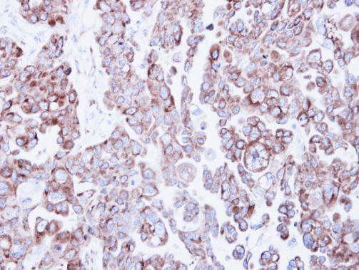
Supportive validation
- Submitted by
- Invitrogen Antibodies (provider)
- Main image

- Experimental details
- Immunohistochemical analysis of paraffin-embedded OVCAR3 xenograft, using Ribosome binding protein 1 (Product # PA5-21392) antibody at 1:500 dilution. Antigen Retrieval: EDTA based buffer, pH 8.0, 15 min.