 Explore
 Validate
 Learn
Learn14-9778-37
antibody from Invitrogen Antibodies
Targeting: LAMP5
BAD-LAMP, C20orf103, dJ1119D9.3, UNC-46
 Western blot
 Immunohistochemistry
Antibody data
- Antibody Data
- Antigen structure
- References [2]
- Comments [0]
- Validations
- Immunohistochemistry [1]
- Other assay [2]
Submit
Validation data
Reference
Comment
Report error
- Product number
- 14-9778-37 - Provider product page

- Provider
- Invitrogen Antibodies
- Product name
- LAMP5 (BAD-LAMP) Monoclonal Antibody (34.2), eBioscience™
- Antibody type
- Monoclonal
- Antigen
- Other
- Description
- The monoclonal antibody 34.2 recognizes human lysosome-associated membrane protein 5 (LAMP5), a member of the LAMP family. LAMP5 differs from other LAMP family members in that its expression is more restricted and is referred to as brain and dendritic cell-associated LAMP-like molecule (BAD-LAMP). In humans, BAD-LAMP is expressed in post-natal neurons and non-activated plasmacytoid dendritic cells. Expression in neurons is localized to intracellular vesicles distributed in specific microdomains along neurites and may play a role in endocytosis. BAD-LAMP is localized to the endoplasmic reticulum-Golgi intermediate compartment (ERGIC) of plasmacytoid dendritic cells and is lost upon TLR activation. This 34.2 antibody has been tested by immunohistochemistry of formalin-fixed paraffin embedded tissue using high or low pH antigen retrieval and can be used at less than or equal to 1 µg/mL. This 34.2 antibody has been tested by western blot analysis of reduced SK-N-SH cell lysates and can be used at less than or equal to 5 µg/mL. It is recommended that the antibody be carefully titrated for optimal performance in the assay of interest. Purity: Greater than 90%, as determined by SDS-PAGE. Aggregation: Less than 10%, as determined by HPLC. Filtration: 0.2 µm post-manufacturing filtered.
- Reactivity
- Human
- Host
- Rat
- Isotype
- IgG
- Antibody clone number
- 34.2
- Vial size
- 2 mg
- Concentration
- 0.5 mg/mL
- Storage
- 4°C
Submitted references BAD-LAMP controls TLR9 trafficking and signalling in human plasmacytoid dendritic cells.
[Development of EEG changes in subacute leukoencephalitis].
Combes A, Camosseto V, N'Guessan P, Argüello RJ, Mussard J, Caux C, Bendriss-Vermare N, Pierre P, Gatti E
Nature communications 2017 Oct 13;8(1):913
Nature communications 2017 Oct 13;8(1):913
[Development of EEG changes in subacute leukoencephalitis].
Sofijanov N, Dukovski M, Sadikario A
Godisen zbornik na Medicinskiot fakultet vo Skopje 1977;23:729-38
Godisen zbornik na Medicinskiot fakultet vo Skopje 1977;23:729-38
No comments: Submit comment
Supportive validation
- Submitted by
- Invitrogen Antibodies (provider)
- Main image
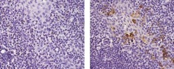
- Experimental details
- Immunohistochemistry of formalin-fixed paraffin embedded human tonsil using 1 µg/mL of Rat IgG1 K Isotype Control Purified (left) or 1 µg/mL of Anti-Human LAMP5 (BAD-LAMP) Purified (right) followed by Anti-Rat IgG Biotin, Avidin HRP, and DAB visualization.Nuclei are counterstained with hematoxylin.
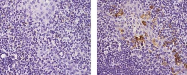
Supportive validation
- Submitted by
- Invitrogen Antibodies (provider)
- Main image
- Experimental details
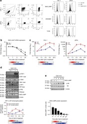
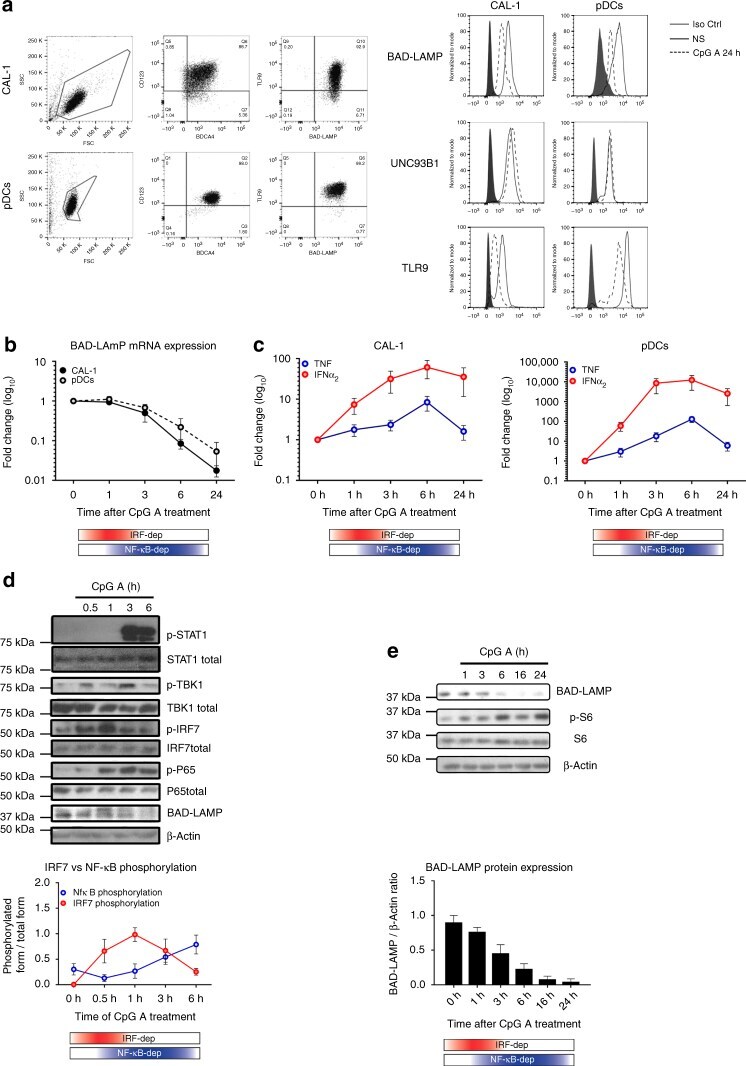
- Fig. 1 BAD-LAMP is down-modulated after IRF7 activation. a ( left ) Flow cytometry analysis of CAL-1 cells ( top ) and freshly isolated pDCs ( bottom ) from healthy donors stained for both extracellular (CD123 and BADCA4) and intracellular (TLR9 and BAD-LAMP) pDCs markers. ( right ) Flow cytometry histogram plots of intracellular staining for BAD-LAMP, TLR9 and UNC93B1 in both CAL-1 ( left ) and freshly isolated pDCs ( right ) at steady state ( black line ) and after 24 h of CpG-A stimulation ( dashed line ). Full grey histograms represent isotype controls staining. Data are representative of a minimum of three independent experiments. b CAL-1 ( black line ) and freshly isolated pDCs ( dashed line ) were treated with CpG-A for indicated times. BAD-LAMP mRNA levels were measured by RT-qPCR. Raw data have been normalised to housekeeping gene (GAPDH) and graphics represent fold change +- s.d. compared to non-stimulated cells from a minimum of three independent experiments. c IFNalpha 2 ( red ) and TNF ( blue ) mRNA level from CAL-1 ( left ) and freshly isolated pDCs ( right ) were measured by RT-qPCR. Raw data have been normalised to housekeeping gene (GAPDH) and graphics represent fold change +- s.d. compared to non-stimulated cells from a minimum of three independent experiments. d ( top ) CAL-1 cells were treated with CpG-A for indicated times prior lysis and sodium dodecyl sulphate-polyacrylamide gel electrophoresis treatment. Expression of BAD-LAMP, TBK1, IRF7, STAT1, the p
- Submitted by
- Invitrogen Antibodies (provider)
- Main image
- Experimental details
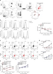
- Fig. 10 BAD-LAMP expression in tumour-associated pDCs correlates with low IFN-alpha production. a ( left ) Flow cytometry pDC gating strategy on both PBMC ( top ) and tumour ( bottom ) is shown. pDCs (BDCA2 + ; CD123 + ) were segregated from conventional dendritic cells (Lin - , MHC II high ) after identification of live hematopoietic cells (CD45 + Live Dead - ). ( right ) CD123 + /BDCA4 + pDCs from blood ( black ) or from primary breast tumours ( red ) were further analysed for BAD-LAMP and MHC II expression. Data are representative of three patients. b Freshly isolated pDCs from healthy donors were treated for 16 h with tumour supernatant either devoid of (snTUM-) or enriched in (snTUM+) TNF and TGF-beta. ( left ) BAD-LAMP and MHC II histogram plots from FACS staining at steady state ( black line ) or 24 h ( dashed line ) after CpG-A treatment. Full grey histogram represents isotype control. Data are representative for five independent experiments performed with eight different tumour supernatants. ( right ) Levels of BAD-LAMP shown as MFI from FACS intracellular staining in pDCs pre-treated with snTUM- ( dashed line ), snTUM + ( red line ) or with medium ( black lines ). MFI +- s.d. from five independent experiments. c Freshly isolated pDCs from heathy donors were treated for 16 h with tumour supernatant either devoid of (snTUM-; black) or enriched in (snTUM + ; red) in TNF and TGF-beta. 2D FACS analysis of intracellular staining for IFN-alpha and TNF cytokines at differen
