Explore
 Validate
 Learn
Learn46-5889-41
antibody from Invitrogen Antibodies
Targeting: ICOSLG
B7-H2, B7h, B7H2, B7RP-1, B7RP1, CD275, GL50, ICOS-L, ICOSL, KIAA0653
 Flow cytometry
Antibody data
- Antibody Data
- Antigen structure
- References [1]
- Comments [0]
- Validations
- Flow cytometry [1]
- Other assay [1]
Submit
Validation data
Reference
Comment
Report error
- Product number
- 46-5889-41 - Provider product page

- Provider
- Invitrogen Antibodies
- Product name
- CD275 (B7-H2) Monoclonal Antibody (MIH12), PerCP-eFluor™ 710, eBioscience™
- Antibody type
- Monoclonal
- Antigen
- Other
- Description
- Description: The MIH12 monoclonal antibody reacts with human B7RP-1, also known as B7h, B7-H2, GL50 and ICOS Ligand. B7RP-1, a member of the B7 family, has a predicted molecular weight of approximately 40 kDa and belongs to the Ig superfamily. Human B7RP-1 is expressed by activated monocytes/macrophages. B7RP-1 binds to the ICOS molecule (AILIM, CRP-1) expressed by activated T cells. The interaction of ICOS/B7RP-1 plays an important role in the T cell costimulation pathway.
- Antibody clone number
- MIH12
- Concentration
- 5 µL/Test
Submitted references Cytomegalovirus restricts ICOSL expression on antigen-presenting cells disabling T cell co-stimulation and contributing to immune evasion.
Angulo G, Zeleznjak J, Martínez-Vicente P, Puñet-Ortiz J, Hengel H, Messerle M, Oxenius A, Jonjic S, Krmpotić A, Engel P, Angulo A
eLife 2021 Jan 18;10
eLife 2021 Jan 18;10
No comments: Submit comment
Supportive validation
- Submitted by
- Invitrogen Antibodies (provider)
- Main image
- Experimental details
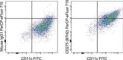
- Normal human peripheral blood monocyte-derived dendritic cells were stained with CD11c Monoclonal Antibody, FITC (Product # 11-0116-42) and Mouse IgG1 kappa Isotype Control, PerCP-eFluor 710 (Product # 46-4714-82) (left) or CD275 (B7H2) Monoclonal Antibody, PerCP-eFluor 710 (right). Total viable cells were used for analysis.

Supportive validation
- Submitted by
- Invitrogen Antibodies (provider)
- Main image
- Experimental details
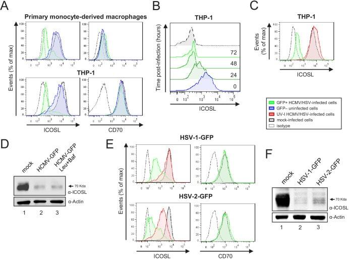
- Figure 11. HCMV, HSV-1, and HSV-2 also limit cell surface expression of ICOSL on antigen-presenting cells (APCs). ( A ) Primary monocyte-derived macrophages (upper panels) and PMA-treated THP-1 cells (bottom panels) were mock-infected or infected for 72 hr with HCMV-GFP at an moi of 10 and analyzed by flow cytometry for cell-surface expression of human ICOSL or CD70 using specific mAbs against each of these receptors. Gray histograms represent the expression of mock-infected cells, green histograms represent the expression on HCMV-infected (GFP+) cells, and blue histograms represent the expression on uninfected (GFP-) cells from the same culture. ( B ) PMA-treated THP-1 cells were mock-infected (time 0) or infected with HCMV-GFP as in A and analyzed by flow cytometry for surface expression of ICOSL at the different time points after infection indicated. ( C ) Same as in A, except that an moi of 20 was used, and THP-1 cells were also exposed for 72 hr to the same amount of HCMV-GFP UV-inactivated (red histogram). ( D ) Equal amounts of lysates from PMA-treated THP-1 cells mock-infected (lane 1) or infected for 72 hr at an moi of 20 with HCMV-GFP (lanes 2 and 3), and when indicated, treated with 250 muM leupeptin and 20 nM of bafilomycin A1 (lane 3), were lysed and analyzed by western blot with antibodies against ICOSL and actin, followed by anti-rabbit IgG-HRP (ICOSL) or anti-mouse IgG-HRP (actin). ( E ) PMA-treated THP-1 cells were mock-infected or infected with HSV-1-GFP, HS