 Explore
 Validate
 Learn
Learn Flow cytometry
Antibody data
- Antibody Data
- Antigen structure
- References [2]
- Comments [0]
- Validations
- Flow cytometry [1]
Submit
Validation data
Reference
Comment
Report error
- Product number
- FAB1139F-025 - Provider product page

- Provider
- R&D Systems
- Product name
- Human Siglec-9 Fluorescein-conjugated Antibody
- Antibody type
- Monoclonal
- Description
- Protein A or G purified from hybridoma culture supernatant. Detects human Siglec-9 in ELISAs and Western blots. In Western blots, no cross-reactivity with recombinant human Siglec-2, -3, -5, -7 or -10 is observed.
- Reactivity
- Human
- Host
- Mouse
- Antigen sequence
Q9Y336- Isotype
- IgG
- Antibody clone number
- 191240
- Vial size
- 25 Tests
- Storage
- Protect from light. Do not freeze. 12 months from date of receipt, 2 to 8 °C as supplied.
Submitted references Increased expression of Siglec-9 in chronic obstructive pulmonary disease.
Structural characterisation of high affinity Siglec-2 (CD22) ligands in complex with whole Burkitt's lymphoma (BL) Daudi cells by NMR spectroscopy.
Zeng Z, Li M, Wang M, Wu X, Li Q, Ning Q, Zhao J, Xu Y, Xie J
Scientific reports 2017 Aug 31;7(1):10116
Scientific reports 2017 Aug 31;7(1):10116
Structural characterisation of high affinity Siglec-2 (CD22) ligands in complex with whole Burkitt's lymphoma (BL) Daudi cells by NMR spectroscopy.
Madge PD, Maggioni A, Pascolutti M, Amin M, Waespy M, Bellette B, Thomson RJ, Kelm S, von Itzstein M, Haselhorst T
Scientific reports 2016 Nov 3;6:36012
Scientific reports 2016 Nov 3;6:36012
No comments: Submit comment
Supportive validation
- Submitted by
- R&D Systems (provider)
- Main image
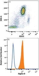
- Experimental details
- Detection of Siglec-9 in Human Blood Granulocytes by Flow Cytometry. Human peripheral blood granulocytes were stained with (A) Mouse Anti-Human Siglec-3/CD33 PE-conjugated Monoclonal Antibody (Catalog # FAB1137P) and (B) Mouse Anti-Human Siglec-9 Fluorescein-conjugated Monoclonal Antibody (Catalog # FAB1139F, filled histogram) or isotype control antibody (Catalog # IC003F, open histogram). View our protocol for Staining Membrane-associated Proteins.
