Explore
 Validate
 Learn
Learn Western blot
 Immunocytochemistry
Antibody data
- Antibody Data
- Antigen structure
- References [0]
- Comments [0]
- Validations
- Immunocytochemistry [1]
- Flow cytometry [3]
Submit
Validation data
Reference
Comment
Report error
- Product number
- 711505 - Provider product page

- Provider
- Invitrogen Antibodies
- Product name
- eIF4A1 Recombinant Polyclonal Antibody
- Antibody type
- Polyclonal
- Antigen
- Synthetic peptide
- Reactivity
- Human, Mouse
- Host
- Rabbit
- Isotype
- IgG
- Vial size
- 100 µg
- Concentration
- 0.5 mg/mL
- Storage
- Store at 4°C short term. For long term storage, store at -20°C, avoiding freeze/thaw cycles.
No comments: Submit comment
Supportive validation
- Submitted by
- Invitrogen Antibodies (provider)
- Main image
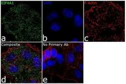
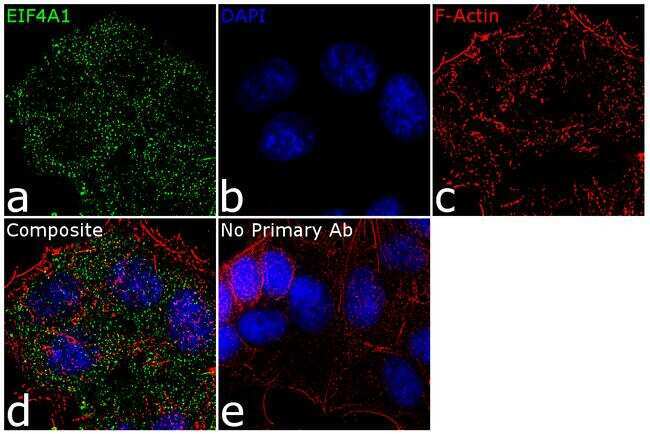
- Experimental details
- For immunofluorescence analysis, MCF7 cells were fixed and permeabilized for detection of endogenous EIF4A1 using Anti- EIF4A1 Recombinant Rabbit Polyclonal Antibody (Product # 711505, 2 µg/mL) and labeled with Goat anti-Rabbit IgG (H+L) Superclonal™ Secondary Antibody, Alexa Fluor® 488 conjugate (Product # A27034, 1:2000). Panel a) shows representative cells that were stained for detection and localization of EIF4A1 protein (green), Panel b) is stained for nuclei (blue) using SlowFade® Gold Antifade Mountant with DAPI (Product # S36938). Panel c) represents cytoskeletal F-actin staining using Rhodamine Phalloidin (Product # R415, 1:300). Panel d) is a composite image of Panels a, b and c clearly demonstrating cytoplasmic localization of EIF4A1. Panel e) represents control cells with no primary antibody to assess background. The images were captured at 60X magnification.
Supportive validation
- Submitted by
- Invitrogen Antibodies (provider)
- Main image
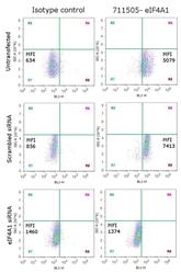
- Experimental details
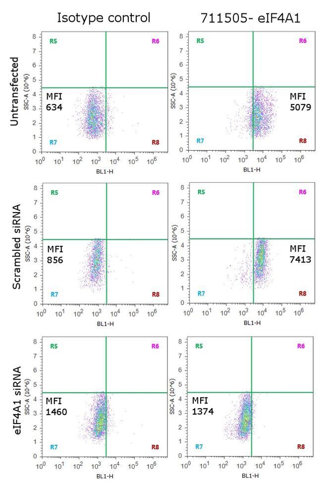
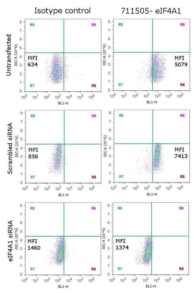
- Knockdown of eIF4A1 was achieved by transfecting HeLa cells with eIF4A1 specific siRNA (Silencer® select Product # s4567). Flow cytometry analysis was performed on cells transfected with eIF4A1 specific siRNA, non-specific scrambled siRNA and untransfected cells. Cells were probed with Anti-eIF4A1 Recombinant Rabbit Polyclonal Antibody (Product # 711505, 5 µg/ 1M cells) or with the matched rabbit isotype control, and detected with Goat anti-Rabbit IgG (H+L) Superclonal™ Secondary Antibody (Alexa Fluor® 488 conjugate, Product # A27034, 0.4 µg/mL, 1:2500). A representative of 10,000 cells were acquired and analyzed for each sample using an Attune® Acoustic Focusing Cytometer (Product # 4468770). No positive events were observed in siRNA transfected cells confirming specificity of the antibody to eIF4A1.
- Submitted by
- Invitrogen Antibodies (provider)
- Main image
- Experimental details
- Knockdown of eIF4A1 was achieved by transfecting HeLa cells with eIF4A1 specific siRNA (Silencer® select Product # s4567). Flow cytometry analysis was performed on cells transfected with eIF4A1 specific siRNA, non-specific scrambled siRNA and untransfected cells. Cells were probed with Anti-eIF4A1 Recombinant Rabbit Polyclonal Antibody (Product # 711505, 5 µg/ 1M cells) or with the matched rabbit isotype control, and detected with Goat anti-Rabbit IgG (H+L) Superclonal™ Secondary Antibody (Alexa Fluor® 488 conjugate, Product # A27034, 0.4 µg/mL, 1:2500). A representative of 10,000 cells were acquired and analyzed for each sample using an Attune® Acoustic Focusing Cytometer (Product # 4468770). No positive events were observed in siRNA transfected cells confirming specificity of the antibody to eIF4A1.
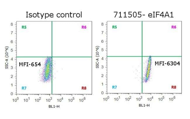
- Submitted by
- Invitrogen Antibodies (provider)
- Main image
- Experimental details
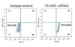
- Flow Cytometry analysis of endogenous eIF4A1 was performed on HeLa cells labeled with Anti-eIF4A1 Recombinant Rabbit Polyclonal Antibody (Product # 711505, 5 µg/ 1M cells) or with rabbit isotype control, and detected with Goat anti-Rabbit IgG (H+L) Superclonal™ Secondary Antibody, (Alexa Fluor® 488 conjugate, Product # A27034, 0.4 µg/mL, 1:2500). A representative of 10,000 cells were acquired and analyzed for each sample using an Attune® Acoustic Focusing Cytometer (Product # 4468770).