Explore
 Validate
 Learn
Learn Western blot
Antibody data
- Antibody Data
- Antigen structure
- References [1]
- Comments [0]
- Validations
- Western blot [1]
- Immunohistochemistry [1]
Submit
Validation data
Reference
Comment
Report error
- Product number
- AF7677 - Provider product page

- Provider
- R&D Systems
- Product name
- Mouse/Rat M-Cadherin/Cadherin-15 Antibody
- Antibody type
- Polyclonal
- Description
- Antigen Affinity-purified. Detects mouse and rat M-Cadherin/Cadherin-15 in direct ELISAs and Western blots. In direct ELISAs, approximately 10% cross-reactivity with recombinant human M-Cadherin/Cadherin-15 is observed, and less than 1% cross-reactivity with recombinant mouse P-Cadherin is observed.
- Reactivity
- Mouse, Rat
- Host
- Sheep
- Conjugate
- Unconjugated
- Antigen sequence
P33146- Isotype
- IgG
- Vial size
- 100 ug
- Concentration
- LYOPH
- Storage
- Use a manual defrost freezer and avoid repeated freeze-thaw cycles. 12 months from date of receipt, -20 to -70 °C as supplied. 1 month, 2 to 8 °C under sterile conditions after reconstitution. 6 months, -20 to -70 °C under sterile conditions after reconstitution.
Submitted references Impairment of cold injury-induced muscle regeneration in mice receiving a combination of bone fracture and alendronate treatment.
Kawada S, Harada A, Hashimoto N
PloS one 2017;12(7):e0181457
PloS one 2017;12(7):e0181457
No comments: Submit comment
Supportive validation
- Submitted by
- R&D Systems (provider)
- Main image
- Experimental details
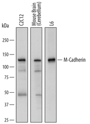
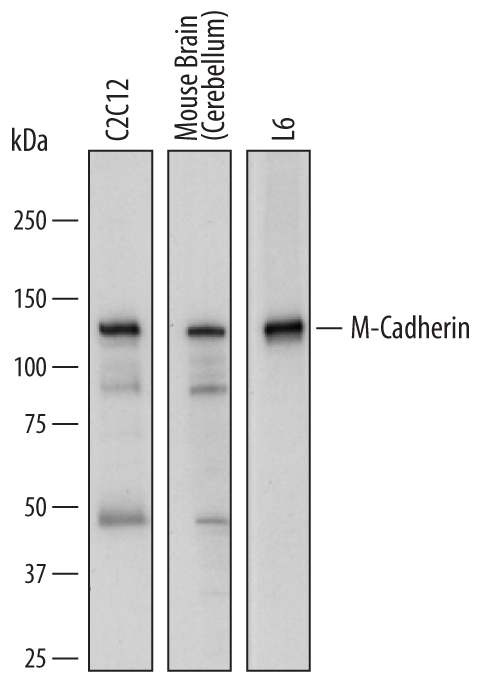
- Detection of Mouse and Rat M-Cadherin/ Cadherin-15 by Western Blot. Western blot shows lysates of C2C12 mouse myoblast cell line, mouse brain (cerebellum) tissue, and L6 rat myoblast cell line. PVDF membrane was probed with 1 µg/mL of Sheep Anti-Mouse/Rat M-Cadherin/Cadherin-15 Antigen Affinity-purified Polyclonal Antibody (Catalog # AF7677) followed by HRP-conjugated Anti-Sheep IgG Secondary Antibody (Catalog # HAF016). A specific band was detected for M-Cadherin/Cadherin-15 at approximately 125 kDa (as indicated). This experiment was conducted under reducing conditions and using Immunoblot Buffer Group 1.
Supportive validation
- Submitted by
- R&D Systems (provider)
- Main image
- Experimental details
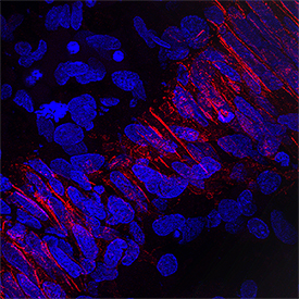
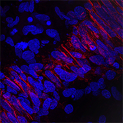
- M-Cadherin/Cadherin-15 in Human Embryonic Muscle. M-Cadherin/Cadherin-15 was detected in perfusion fixed frozen sections of human embryonic muscle using Sheep Anti-Mouse/Rat M-Cadherin/Cadherin-15 Antigen Affinity-purified Polyclonal Antibody (Catalog # AF7677) at 1.7 µg/mL overnight at 4 °C. Tissue was stained using the NorthernLights™ 557-conjugated Anti-Sheep IgG Secondary Antibody (red; Catalog # NL010) and counterstained with DAPI (blue). Specific staining was localized to plasma membrane. View our protocol for Fluorescent IHC Staining of Frozen Tissue Sections.