 Explore
 Validate
 Learn
Learn Western blot
Antibody data
- Antibody Data
- Antigen structure
- References [2]
- Comments [0]
- Validations
- Western blot [1]
- Immunocytochemistry [1]
- Flow cytometry [1]
Submit
Validation data
Reference
Comment
Report error
- Product number
- AF4096 - Provider product page

- Provider
- R&D Systems
- Product name
- Human M-Cadherin/Cadherin-15 Antibody
- Antibody type
- Polyclonal
- Description
- Antigen Affinity-purified. Detects human M-Cadherin/Cadherin-15 in direct ELISAs and Western blots.
- Reactivity
- Human
- Host
- Sheep
- Conjugate
- Unconjugated
- Antigen sequence
P55291- Isotype
- IgG
- Vial size
- 100 ug
- Concentration
- LYOPH
- Storage
- Use a manual defrost freezer and avoid repeated freeze-thaw cycles. 12 months from date of receipt, -20 to -70 °C as supplied. 1 month, 2 to 8 °C under sterile conditions after reconstitution. 6 months, -20 to -70 °C under sterile conditions after reconstitution.
Submitted references Interleukin-1beta (IL-1β)-induced Notch ligand Jagged1 suppresses mitogenic action of IL-1β on human dystrophic myogenic cells.
Homeostatic regulation of T cell trafficking by a B cell-derived peptide is impaired in autoimmune and chronic inflammatory disease.
Nagata Y, Kiyono T, Okamura K, Goto YI, Matsuo M, Ikemoto-Uezumi M, Hashimoto N
PloS one 2017;12(12):e0188821
PloS one 2017;12(12):e0188821
Homeostatic regulation of T cell trafficking by a B cell-derived peptide is impaired in autoimmune and chronic inflammatory disease.
Chimen M, McGettrick HM, Apta B, Kuravi SJ, Yates CM, Kennedy A, Odedra A, Alassiri M, Harrison M, Martin A, Barone F, Nayar S, Hitchcock JR, Cunningham AF, Raza K, Filer A, Copland DA, Dick AD, Robinson J, Kalia N, Walker LSK, Buckley CD, Nash GB, Narendran P, Rainger GE
Nature medicine 2015 May;21(5):467-475
Nature medicine 2015 May;21(5):467-475
No comments: Submit comment
Supportive validation
- Submitted by
- R&D Systems (provider)
- Main image

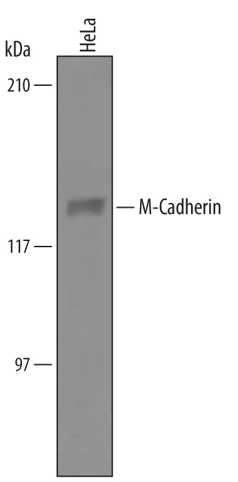
- Experimental details
- Detection of Human M-Cadherin/Cadherin-15 by Western Blot. Western blot shows lysates of HeLa human cervical epithelial carcinoma cell line. PVDF Membrane was probed with 1 µg/mL of Sheep Anti-Human M-Cadherin/Cadherin-15 Antigen Affinity-purified Polyclonal Antibody (Catalog # AF4096) followed by HRP-conjugated Anti-Sheep IgG Secondary Antibody (Catalog # HAF016). A specific band was detected for M-Cadherin/Cadherin-15 at approximately 130 kDa (as indicated). This experiment was conducted under reducing conditions and using Immunoblot Buffer Group 8.
Supportive validation
- Submitted by
- R&D Systems (provider)
- Main image
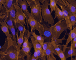
- Experimental details
- M-Cadherin/Cadherin-15 in C2C12 Mouse Cell Line. M-Cadherin/Cadherin-15 was detected in immersion fixed C2C12 mouse myoblast cell line using Sheep Anti-Human M-Cadherin/Cadherin-15 Antigen Affinity-purified Polyclonal Antibody (Catalog # AF4096) at 10 µg/mL for 3 hours at room temperature. Cells were stained using the Northern-Lights™ 557-conjugated Anti-Mouse IgG Secondary Antibody (red; Catalog # NL007) and counter-stained with DAPI (blue). Specific staining was localized to cell surfaces and cytoplasm. View our protocol for Fluorescent ICC Staining of Cells on Coverslips.

Supportive validation
- Submitted by
- R&D Systems (provider)
- Main image
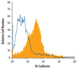
- Experimental details
- Detection of M-Cadherin/ Cadherin-15 in C2C12 Mouse Cell Line by Flow Cytometry. C2C12 mouse myoblast cell line was stained with Sheep Anti-Human M-Cadherin/ Cadherin-15 Antigen Affinity-purified Polyclonal Antibody (Catalog # AF4096, filled histogram) or control antibody (Catalog # 5-001-A, open histogram), followed by NorthernLights™ 557-conjugated Anti-Sheep IgG Secondary Antibody (Catalog # NL010).
