 Explore
 Validate
 Learn
Learn Immunohistochemistry
Antibody data
- Antibody Data
- Antigen structure
- References [0]
- Comments [0]
- Validations
- Immunohistochemistry [11]
Submit
Validation data
Reference
Comment
Report error
- Product number
- AMAb90854 - Provider product page

- Provider
- Atlas Antibodies
- Proper citation
- Atlas Antibodies Cat#AMAb90854, RRID:AB_2665692
- Product name
- Anti-RHOT1
- Antibody type
- Monoclonal
- Reactivity
- Human
- Host
- Mouse
- Conjugate
- Unconjugated
- Antigen sequence
THIVDYSEAEQSDEQLHQEISQANVICIVYAVNNK
HSIDKVTSRWIPLINERTDKDSRLPLILVGNKSDL
VEYSSMETILPIMNQYTEIE- Epitope
- Binds to an epitope located within the peptide sequence ERTDKDSRLP as determined by overlapping synthetic peptides.
- Isotype
- IgG
- Antibody clone number
- CL1095
- Vial size
- 100 µl
- Storage
- Store at +4°C for short term storage. Long time storage is recommended at -20°C.
No comments: Submit comment
Supportive validation
- Submitted by
- Atlas Antibodies (provider)
- Main image
- Experimental details
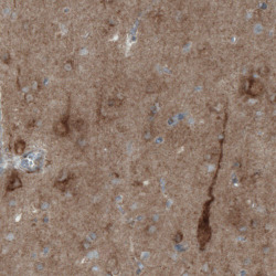
- Immunohistochemical staining of human cerebral cortex shows immunoreactivity in both neuronal cell bodies and neuropil.

- Submitted by
- Atlas Antibodies (provider)
- Main image
- Experimental details
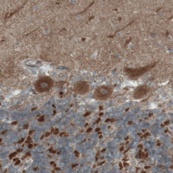
- Immunohistochemical staining of human cerebellum shows strong positivity in Purkinje cells, as well as in molecular and granular cell layers.

- Submitted by
- Atlas Antibodies (provider)
- Main image
- Experimental details
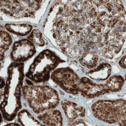
- Immunohistochemical staining of human kidney shows strong immunoreactivity in renal tubules and moderate staining in glomeruli.

- Submitted by
- Atlas Antibodies (provider)
- Main image
- Experimental details
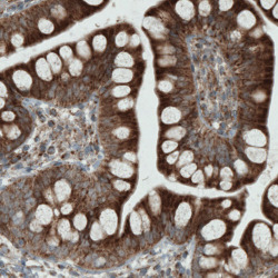
- Immunohistochemical staining of human small intestine shows cytoplasmic immunoreactivity in glandular cells.

- Submitted by
- Atlas Antibodies (provider)
- Main image

- Experimental details
- Immunohistochemical staining of human testis shows strong granular cytoplasmic immunoreactivity in seminiferous tubules cells.

- Submitted by
- Atlas Antibodies (provider)
- Main image
- Experimental details
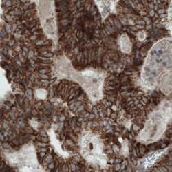
- Immunohistochemical staining of human colorectal cancer shows strong cytoplasmic immunoreactivity in tumor cells.

- Submitted by
- Atlas Antibodies (provider)
- Main image
- Experimental details
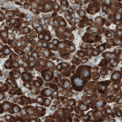
- Immunohistochemical staining of human liver cancer shows strong cytoplasmic positivity in tumor cells.

- Submitted by
- Atlas Antibodies (provider)
- Main image
- Experimental details
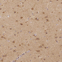
- Immunohistochemical staining of human cerebral cortex shows moderate to strong cytoplasmic positivity in neuronal cells, as well as positivity in neuropil.

- Submitted by
- Atlas Antibodies (provider)
- Main image
- Experimental details
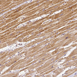
- Immunohistochemical staining of human heart muscle shows moderate cytoplasmic positivity in cardiomyocytes.

- Submitted by
- Atlas Antibodies (provider)
- Main image
- Experimental details
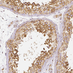
- Immunohistochemical staining of human testis shows moderate cytoplasmic positivity in cells in seminiferous ducts.

- Submitted by
- Atlas Antibodies (provider)
- Main image
- Experimental details
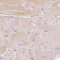
- Immunohistochemical staining of human skeletal muscle shows only very weak cytoplasmic positivity in striated muscle fibers.
