 Explore
 Validate
 Learn
Learn Western blot
 Immunocytochemistry
Antibody data
- Antibody Data
- Antigen structure
- References [1]
- Comments [0]
- Validations
- Western blot [5]
- Immunocytochemistry [1]
- Immunohistochemistry [2]
Submit
Validation data
Reference
Comment
Report error
- Product number
- GTX101676 - Provider product page

- Provider
- GeneTex
- Proper citation
- GeneTex Cat#GTX101676, RRID:AB_1949820
- Product name
- Calnexin antibody [N3C2], Internal
- Antibody type
- Polyclonal
- Reactivity
- Human, Mouse, Rat
- Host
- Rabbit
Submitted references Cellular uptake of extracellular vesicles is mediated by clathrin-independent endocytosis and macropinocytosis.
Costa Verdera H, Gitz-Francois JJ, Schiffelers RM, Vader P
Journal of controlled release : official journal of the Controlled Release Society 2017 Nov 28;266:100-108
Journal of controlled release : official journal of the Controlled Release Society 2017 Nov 28;266:100-108
No comments: Submit comment
Enhanced validation
Supportive validation
- Submitted by
- GeneTex (provider)
- Enhanced method
- Genetic validation
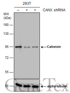
- Main image
- Experimental details
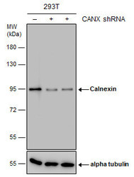
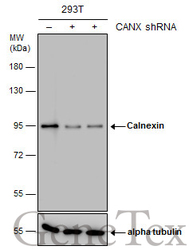
- Non-transfected (¡V) and transfected (+) 293T whole cell extracts (15 ?g) were separated by 7.5% SDS-PAGE, and the membrane was blotted with Calnexin antibody [N3C2], Internal (GTX101676) diluted at 1:10000. The HRP-conjugated anti-rabbit IgG antibody (GTX213110-01) was used to detect the primary antibody.
Supportive validation
- Submitted by
- GeneTex (provider)
- Main image
- Experimental details
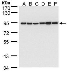
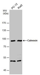
- Sample (30 ?g of whole cell lysate)A: 293TB: A431 (GTX27909)C: H1299D: HeLa S3E: HepG2 (GTX27900)F: Molt-4 (GTX27912)7.5% SDS PAGEGTX101676 diluted at 1:5000The HRP-conjugated anti-rabbit IgG antibody (GTX213110-01) was used to detect the primary antibody.

- Submitted by
- GeneTex (provider)
- Main image
- Experimental details
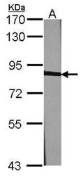
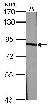
- Sample (30 ?g of whole cell lysate) A:NIH-3T37.5% SDS PAGE GTX101676 diluted at 1:1000 The HRP-conjugated anti-rabbit IgG antibody (GTX213110-01) was used to detect the primary antibody.
- Submitted by
- GeneTex (provider)
- Main image
- Experimental details
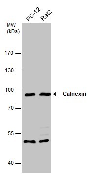
- Various whole cell extracts (30 ?g) were separated by 7.5% SDS-PAGE, and the membrane was blotted with Calnexin antibody (GTX101676) diluted at 1:1000. The HRP-conjugated anti-rabbit IgG antibody (GTX213110-01) was used to detect the primary antibody.
- Submitted by
- GeneTex (provider)
- Main image
- Experimental details
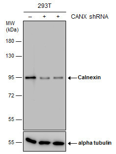
- Non-transfected (¡V) and transfected (+) 293T whole cell extracts (15 ?g) were separated by 7.5% SDS-PAGE, and the membrane was blotted with Calnexin antibody [N3C2], Internal (GTX101676) diluted at 1:10000. The HRP-conjugated anti-rabbit IgG antibody (GTX213110-01) was used to detect the primary antibody.
Supportive validation
- Submitted by
- GeneTex (provider)
- Main image
- Experimental details
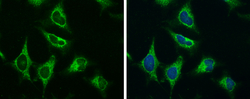
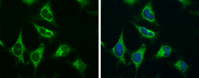
- Calnexin antibody [N3C2], Internal detects Calnexin protein at endoplasmic reticulum by immunofluorescent analysis.Sample: HeLa cells were fixed in ice-cold MeOH for 5 min.Green: Calnexin protein stained by Calnexin antibody [N3C2], Internal (GTX101676) diluted at 1:200.Blue: Hoechst 33342 staining.
Supportive validation
- Submitted by
- GeneTex (provider)
- Main image
- Experimental details
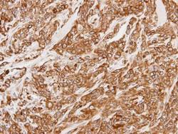
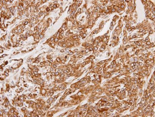
- Immunohistochemical analysis of paraffin-embedded SAS xenograft, using Calnexin(GTX101676) antibody at 1:100 dilution.
- Submitted by
- GeneTex (provider)
- Main image
- Experimental details
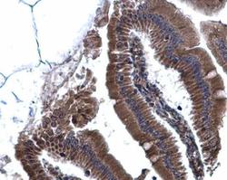
- Calnexin antibody [N3C2], Internal detects Calnexin protein at cytoplasm on mouse duodenum by immunohistochemical analysis. Sample: Paraffin-embedded mouse duodenum. Calnexin antibody [N3C2], Internal (GTX101676) diluted at 1:500.
