Explore
 Validate
 Learn
Learn Immunocytochemistry
Antibody data
- Antibody Data
- Antigen structure
- References [3]
- Comments [0]
- Validations
- Immunocytochemistry [1]
- Other assay [2]
Submit
Validation data
Reference
Comment
Report error
- Product number
- MA3-027-A488 - Provider product page

- Provider
- Invitrogen Antibodies
- Product name
- Calnexin Monoclonal Antibody (AF18), Alexa Fluor™ 488
- Antibody type
- Monoclonal
- Antigen
- Other
- Description
- MA3027A488 detects calnexin from human and mouse tissues. The MA3027A488 antigen is Focus human hepatoma cell lysate.
- Reactivity
- Human, Mouse
- Host
- Mouse
- Conjugate
- Green dye
- Isotype
- IgG
- Antibody clone number
- AF18
- Vial size
- 50 µL
- Concentration
- 1 mg/mL
- Storage
- 4° C, store in dark, DO NOT FREEZE!
Submitted references A key anti-viral protein, RSAD2/VIPERIN, restricts the release of measles virus from infected cells.
Surf4 (Erv29p) binds amino-terminal tripeptide motifs of soluble cargo proteins with different affinities, enabling prioritization of their exit from the endoplasmic reticulum.
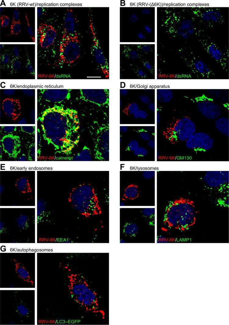
Effects of an In-Frame Deletion of the 6k Gene Locus from the Genome of Ross River Virus.
Kurokawa C, Iankov ID, Galanis E
Virus research 2019 Apr 2;263:145-150
Virus research 2019 Apr 2;263:145-150
Surf4 (Erv29p) binds amino-terminal tripeptide motifs of soluble cargo proteins with different affinities, enabling prioritization of their exit from the endoplasmic reticulum.
Yin Y, Garcia MR, Novak AJ, Saunders AM, Ank RS, Nam AS, Fisher LW
PLoS biology 2018 Aug;16(8):e2005140
PLoS biology 2018 Aug;16(8):e2005140
Effects of an In-Frame Deletion of the 6k Gene Locus from the Genome of Ross River Virus.
Taylor A, Melton JV, Herrero LJ, Thaa B, Karo-Astover L, Gage PW, Nelson MA, Sheng KC, Lidbury BA, Ewart GD, McInerney GM, Merits A, Mahalingam S
Journal of virology 2016 Apr;90(8):4150-4159
Journal of virology 2016 Apr;90(8):4150-4159
No comments: Submit comment
Supportive validation
- Submitted by
- Invitrogen Antibodies (provider)
- Main image

- Experimental details
- Immunofluorescent analysis of Calnexin (green) in HeLa cells. The cells were fixed with 4% Paraformaldehyde in PBS for 15 minutes at room temperature, and blocked with 3% BSA in PBS (Product # 37525) for 30 minutes at room temperature. Cells were stained with a Calnexin Monoclonal Antibody, AlexaFluor 488 conjugate (Product # MA3-027-A488) at a dilution of 5 µg/mL in blocking buffer for 1 hour at room temperature protected from light. Nuclei (blue) were stained with Hoechst Dye (Product # 62249) at a dilution of 1:10,000 in blocking buffer. Images were taken on a Thermo Scientific ToxInsight Instrument at 20X magnification.
- Conjugate
- Green dye

Supportive validation
- Submitted by
- Invitrogen Antibodies (provider)
- Main image
- Experimental details
- NULL
- Conjugate
- Green dye
- Submitted by
- Invitrogen Antibodies (provider)
- Main image
- Experimental details
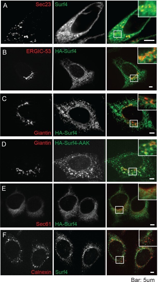
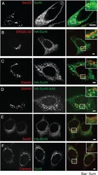
- Fig 5 Immunofluorescence microscopy of HEK293A cells shows Surf4 accumulates in and around ERESs. (A) Fluorescent signal for endogenous Surf4 (green) was strongest at punctate structures positive for ERES marker Sec23 (red). Note additional Surf4 fluorescence in weblike structures surrounding ERES. (B) HA-Surf4 signal (green) was observed within the ERGIC (ERGIC-53, red). (C) HA-Surf4 (green) showed only low levels of colocalization with cis -Golgi marker, giantin (red). (D) Mutation of proposed COPI recycling motif by replacement of two of three near-carboxy-terminal lysines to alanines (HA-Surf4-AAK, green) increased colocalization with cis -Golgi marker, giantin (red). (E) Newly synthesized HA-Surf4 (green) was found at low levels in the rER (Sec61 marker, red). (F) Surf4 (green) did not colocalize with chaperone, calnexin (red), in the quality control domain. HEK293A cells were transfected with wild-type HA-tagged Surf4 plasmid (B, C, E), or Surf4 KO HEK293A cells were transfected with carboxyl-terminal di-lysine mutation, HA- Surf4 -AAK (D), 18 hr prior to fixation. Bars = 5mum. The cells in each panel are shown 3 times, first with organelle marker, then Surf4 and final panel (with magnified insert) showing overlap. Alexa Fluor secondary antibodies were used for detection. Images were obtained using an LSM 780 (Carl Zeiss) confocal microscope (488 and 561 nm excitation lines; 500-560 and 600-660 nm capture) and Zeiss Axio Imager Z1 with Apotome 2 (single Z stac
- Conjugate
- Green dye