Explore
 Validate
 Learn
Learn Western blot
 Immunocytochemistry
 Immunohistochemistry
Antibody data
- Antibody Data
- Antigen structure
- References [2]
- Comments [0]
- Validations
- Immunocytochemistry [4]
- Other assay [2]
Submit
Validation data
Reference
Comment
Report error
- Product number
- PA5-19169 - Provider product page

- Provider
- Invitrogen Antibodies
- Product name
- Calnexin Polyclonal Antibody
- Antibody type
- Polyclonal
- Antigen
- Synthetic peptide
- Description
- This antibody is predicted to react with bovine, canine, porcine and rat based on sequence homology. This antibody is tested in Peptide ELISA: antibody detection limit dilution 128,000.
- Reactivity
- Human
- Host
- Goat
- Isotype
- IgG
- Vial size
- 100 μg
- Concentration
- 0.5 mg/mL
- Storage
- -20°C, Avoid Freeze/Thaw Cycles
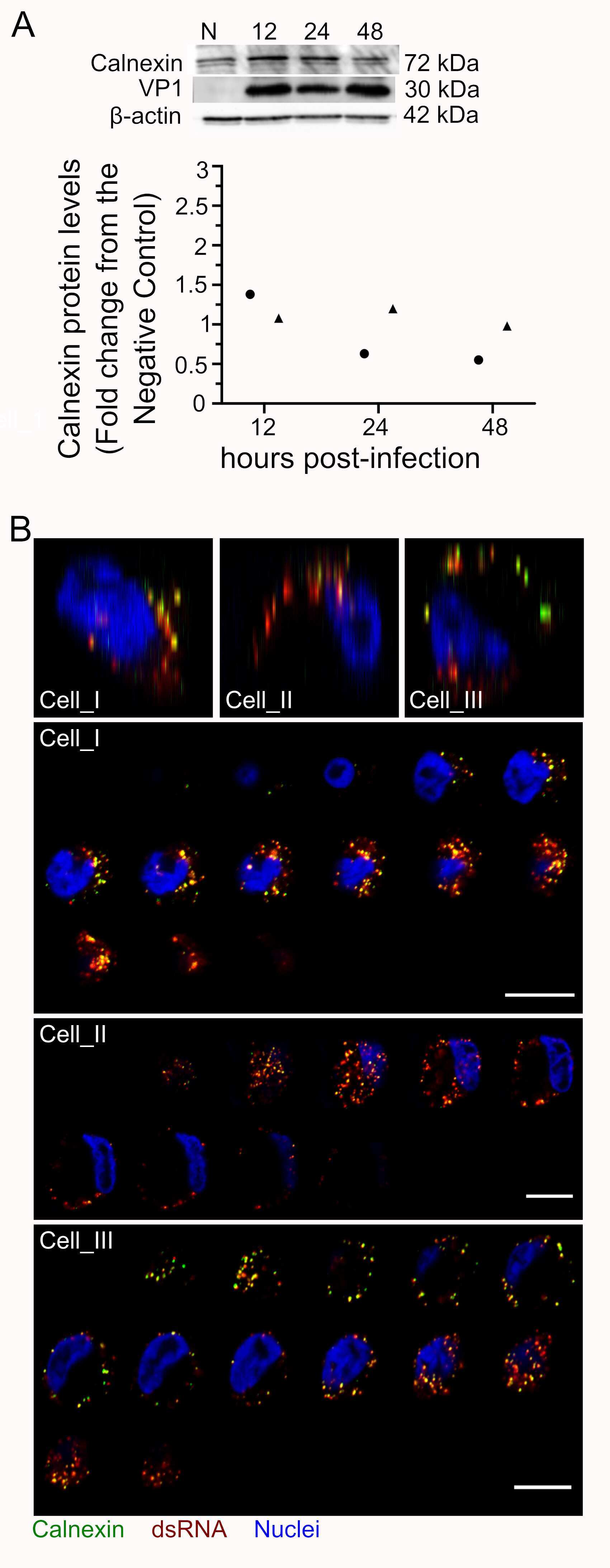
Submitted references Rhinovirus C replication is associated with the endoplasmic reticulum and triggers cytopathic effects in an in vitro model of human airway epithelium.
Ubiquitination of G3BP1 mediates stress granule disassembly in a context-specific manner.
Gagliardi TB, Goldstein ME, Song D, Gray KM, Jung JW, Ignacio MA, Stroka KM, Duncan GA, Scull MA
PLoS pathogens 2022 Jan;18(1):e1010159
PLoS pathogens 2022 Jan;18(1):e1010159
Ubiquitination of G3BP1 mediates stress granule disassembly in a context-specific manner.
Gwon Y, Maxwell BA, Kolaitis RM, Zhang P, Kim HJ, Taylor JP
Science (New York, N.Y.) 2021 Jun 25;372(6549):eabf6548
Science (New York, N.Y.) 2021 Jun 25;372(6549):eabf6548
No comments: Submit comment
Supportive validation
- Submitted by
- Invitrogen Antibodies (provider)
- Main image
- Experimental details
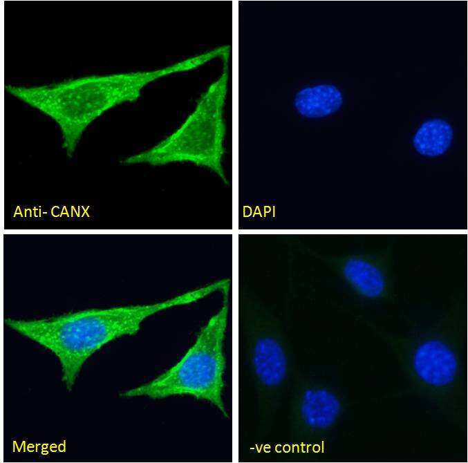
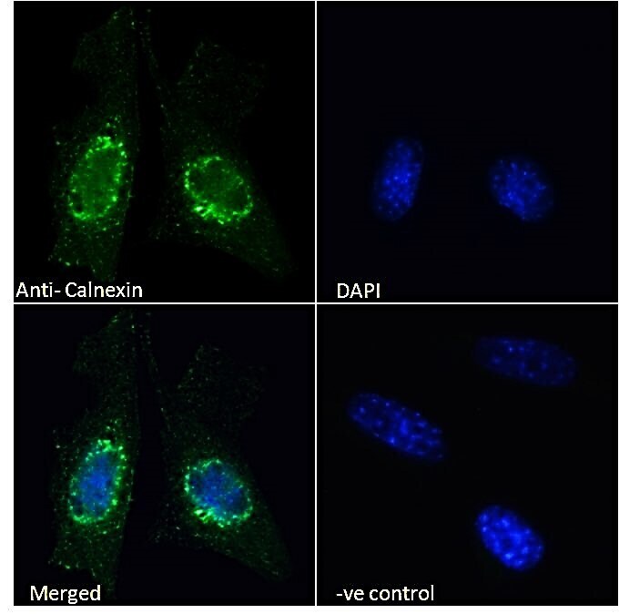
- Immunofluorescence analysis of Calnexin in NIH3T3 cells using a Calnexin monoclonal antibody (Product # PA5-19169) at 5 µg/mL for1hr. The cells were paraformaldehyde fixed and permeabilized with 0.15% Triton. Primary incubation was followed by Alexa Fluor 488 secondary antibody (2 µg/mL) showing cytoplasmic staining. The nuclear stain is DAPI (blue). Negative control: Unimmunized goat IgG (5 µg/mL)followed by Alexa Fluor 488 secondary antibody (2 µg/mL).
- Submitted by
- Invitrogen Antibodies (provider)
- Main image
- Experimental details
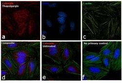
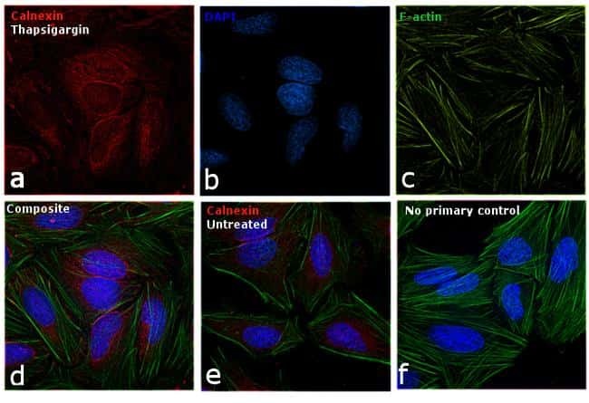
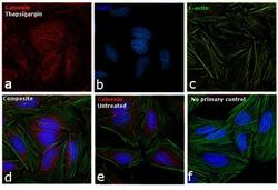
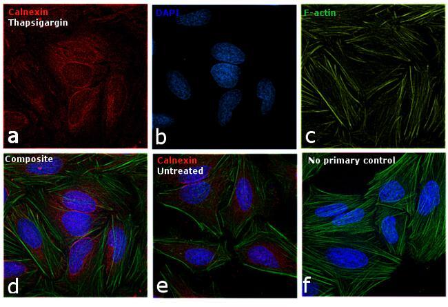
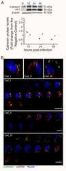
- Immunofluorescence analysis of Calnexin was performed using 70% confluent log phase HeLa cells treated with Thapsigargin (1uM for 24hrs). The cells were fixed with 4% paraformaldehyde for 10 minutes, permeabilized with 0.1% Triton™ X-100 for 15 minutes, and blocked with 1% BSA for 1 hour at room temperature. The cells were labeled with Calnexin Polyclonal Antibody (Product # PA5-19169) at 1:200 dilution in 0.1% BSA, incubated at 4 degree celsius overnight and then labeled with Rabbit anti-Goat IgG (H+L) Superclonal™ Secondary Antibody, Alexa Fluor 594 conjugate (Product # A27016) at a dilution of 1:2000 for 45 minutes at room temperature (Panel a: red). Nuclei (Panel b: blue) were stained with ProLong™ Diamond Antifade Mountant with DAPI (Product # P36962). F-actin (Panel c: green) was stained with Alexa Fluor™ 488 Phalloidin (Product # A12379, 1:300). Panel d represents the merged image showing Calnexin in the ER and cytoplasm. Panel e represents the untreated cells showing lower expression levels. Panel f represents control cells with no primary antibody to assess background. The images were captured at 60X magnification.
- Submitted by
- Invitrogen Antibodies (provider)
- Main image
- Experimental details
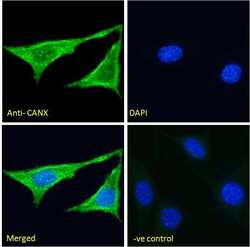
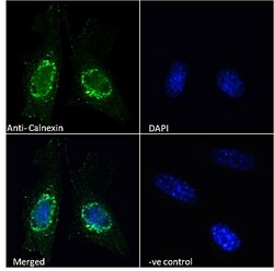
- Immunocytochemistry analysis of Calnexin using Calnexin Polyclonal Antibody (Product # PA5-19169) in paraformaldehyde fixed NIH3T3 cells, permeabilized with 0.15% Triton. Primary incubation 1hr (10 µg/mL) followed by Alexa Fluor 488 secondary antibody (2 µg/mL), showing endoplasmic reticulum staining. The nuclear stain is DAPI (blue). Negative control: Unimmunized goat IgG (10 µg/mL) followed by Alexa Fluor 488 secondary antibody (2 µg/mL).
Supportive validation
- Submitted by
- Invitrogen Antibodies (provider)
- Main image
- Experimental details
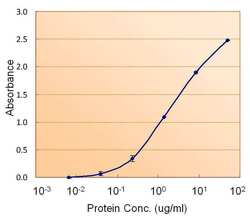
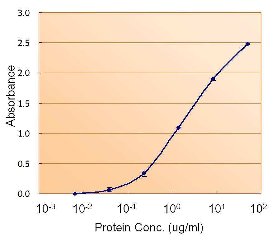
- ELISA of Calnexin using two Calnexin antibodies. The reporter antibody (Product # PA5-19169) was used at a concentration of 1.5 µg/ml and the reporter antibody was used at a concentration of 2.5 µg/ml.
- Submitted by
- Invitrogen Antibodies (provider)
- Main image
- Experimental details
- NULL