 Explore
 Validate
 Learn
Learn Western blot
 Immunohistochemistry
Antibody data
- Antibody Data
- Antigen structure
- References [2]
- Comments [0]
- Validations
- Western blot [1]
- Immunocytochemistry [1]
Submit
Validation data
Reference
Comment
Report error
- Product number
- AB0037-200 - Provider product page

- Provider
- SICGEN
- Proper citation
- SICGEN Cat#AB0037-200, RRID:AB_2333117
- Product name
- Anti-CANX
- Antibody type
- Polyclonal
- Description
- Goat polyclonal to CANX (Calnexin) - endoplasmic reticulum (ER) membrane marker. CANX is a member of the Calnexin family of molecular chaperones. This protein is a calcium-binding, ER-associated protein that interacts transiently with newly synthesized N-linked glycoproteins, facilitating protein folding and assembly. It may also play a central role in the quality control of protein folding by retaining incorrectly folded protein subunits within the ER for degradation.
- Reactivity
- Human, Mouse, Rat, Bovine, Canine, Chicken/Avian, Donkey, Feline, Goat, Guinea Pig, Hamster, Horse, Porcine, Rabbit, Sheep, Simian, Other
- Host
- Goat
- Conjugate
- Unconjugated
- Isotype
- IgG
- Vial size
- 400 µg
- Concentration
- 2 mg/ml
- Storage
- Store at -20 C for long-term storage. Store at 2-8 C for up to one month.
- Handling
- The antibody solution should be gently mixed before use.
Submitted references Methylglyoxal-induced glycation changes adipose tissue vascular architecture, flow and expansion, leading to insulin resistance.
Host cell autophagy contributes to Plasmodium liver development.
Rodrigues T, Matafome P, Sereno J, Almeida J, Castelhano J, Gamas L, Neves C, Gonçalves S, Carvalho C, Arslanagic A, Wilcken E, Fonseca R, Simões I, Conde SV, Castelo-Branco M, Seiça R
Scientific reports 2017 May 10;7(1):1698
Scientific reports 2017 May 10;7(1):1698
Host cell autophagy contributes to Plasmodium liver development.
Thieleke-Matos C, Lopes da Silva M, Cabrita-Santos L, Portal MD, Rodrigues IP, Zuzarte-Luis V, Ramalho JS, Futter CE, Mota MM, Barral DC, Seabra MC
Cellular microbiology 2016 Mar;18(3):437-50
Cellular microbiology 2016 Mar;18(3):437-50
No comments: Submit comment
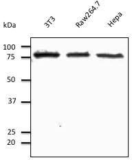
Supportive validation
- Submitted by
- SICGEN (provider)
- Main image
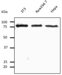
- Experimental details
- Lysates at 50 µg per lane
- Sample type
- 3T3, Raw264.7, Hepa1-6
- Primary Ab dilution
- 1/500
- Conjugate
- Horseradish Peroxidase
- Secondary Ab
- Secondary Ab
- Secondary Ab dilution
- 1/10,000
Supportive validation
- Submitted by
- SICGEN (provider)
- Main image
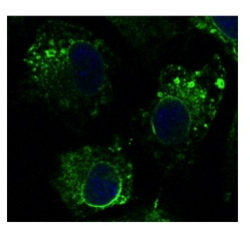
- Experimental details
- Cells fixed with 4% of PFA
- Sample type
- Hepa1-6
- Primary Ab dilution
- 1/100
- Conjugate
- Green dye
